
Brain tumor detection system based on magnetic resonance electrical impedance tomography

A technology of electrical impedance imaging and brain tumors, which is applied in the field of biomedical imaging, can solve the problems of low precision and low image resolution, and achieve the effects of improving morbidity, improving reconstruction quality, and enriching information

Pending Publication Date: 2017-08-22
CHANGSHA UNIVERSITY OF SCIENCE AND TECHNOLOGY
View PDF2 Cites 2 Cited by
  • Summary
  • Abstract
  • Description
  • Claims
  • Application Information

AI Technical Summary

Problems solved by technology

[0010] The purpose of the present invention is to provide a brain tumor detection system based on magnetic resonance electrical impedance imaging, which solves the problems of low image resolution and low precision in the background technology. See the description below for details

Method used


Image

  • Brain tumor detection system based on magnetic resonance electrical impedance tomography
  • Brain tumor detection system based on magnetic resonance electrical impedance tomography
  • Brain tumor detection system based on magnetic resonance electrical impedance tomography

Examples


Embodiment 1

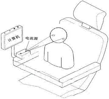
[0035] Embodiment 1: see image 3 , the stimulus source in the brain tumor detection system is a current source with a certain frequency and intensity. The current excitation signal is stimulated to the measured object through the electrode array pasted around the human brain, and the brain will generate a magnetic field; the magnetic field information at different positions can be collected through the PCI bus-based data acquisition card placed around the measured object; Finally, the image reconstruction algorithm is used to obtain the distribution image of the patient's brain tumor area on the computer. When performing brain tumor detection, the subject sits on a chair, places the excitation electrodes on the brain of the imaging body, connects the data acquisition card based on the PCI bus to the detection chair through a bracket of a certain shape, and fixes it on the chair. Around the brain of the imaging subject, the excitation and measurement process is controlled by ...

Embodiment 2

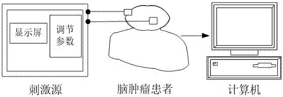
[0036] Example 2: see Figure 4, the excitation source in the brain tumor detection system is a voltage source with a certain frequency and intensity. The current excitation signal is stimulated to the measured object through the electrode array pasted around the human brain, and the brain will generate a magnetic field; the magnetic field information at different positions can be collected through the PCI bus-based data acquisition card placed around the measured object; Finally, the image reconstruction algorithm is used to obtain the distribution image of the patient's brain tumor area on the computer. When performing brain tumor detection, the subject lies flat on the detection bed, places the excitation electrodes on the brain of the imaging body, connects the data acquisition card based on the PCI bus to the detection bed through a bracket of a certain shape, and fixes it on the detection bed. Around the brain of the imaging subject, the excitation and measurement proce...


PUM


Abstract

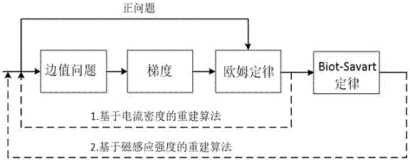
The invention discloses a brain tumor detection system based on magnetic resonance electrical impedance tomography. A stimulus system injects a safe exciting current of a certain frequency to the brain of an imaging body through a stimulus electrode attached to the tested imaging body; an information collection module is composed of data acquisition cards based on a PCI bus and used for collecting magnetic field intensity of the brain, wherein the magnetic field intensity is generated due to the injected exciting current; a software control module is used for controlling current source excitation and the magnetic field collection process in the whole brain tumor detection process; an image reconstruction module acquires the image of brain tumor electrical impedance distribution based on an electrical impedance image reconstruction algorithm. According to the system, the magnetic field information of the brain is collected through the data acquisition cards based on the PCI bus, the amount of information is enlarged, the ill state of image reconstruction with traditional electrical impedance tomography systems is changed, and the resolution of electrical impedance (or impedance spectroscopy) tomography is improved; moreover, the detection system is high in detection precision, high in reliability and harmless to human bodies, and the detection system has good application value for clinical detection and diagnosis of brain tumors.

Description

technical field [0001] The invention relates to the field of biomedical imaging, in particular to a brain tumor detection system based on magnetic resonance electrical impedance imaging. Background technique [0002] Electrical impedance tomography is a non-invasive, precise and functional imaging modality for the detection of brain tumors. patent [1] The head model is processed in layers, according to the prior information of the spatial distribution, the skull artifact is reduced, and the target in the cranial cavity is highlighted, which has the advantages of fast speed and no damage to the human body. In addition, the brain electrical impedance imaging monitoring system improved by the Fourth Military Medical University [2] , through the improvement and selection of the electrode structure, the anti-noise ability of the electrode is improved, the convenience of electrode operation is enhanced, and each functional module of the system is improved. The research results ...

Claims


Application Information

Patent Timeline
IPC IPC(8): A61B5/053A61B5/055
CPCA61B5/053A61B5/055A61B5/4064
Inventor 韩立哲尹剑利沈文旗魏晓英张彦涛邓友均朱抗刘佳彭超敏
Owner CHANGSHA UNIVERSITY OF SCIENCE AND TECHNOLOGY
Learn More