CT digital subtraction angiography (CT/DSA) imaging system and method

An angiography and digital subtraction technology, applied in the field of medical X-ray imaging systems, can solve the problems of reduced image quality, unfavorable clinical diagnosis and treatment, complex structure and control, etc., to improve image quality, improve image quality, and eliminate motion artifacts. Effect

Inactive Publication Date: 2017-09-15
NANJING UNIV OF AERONAUTICS & ASTRONAUTICS
View PDF3 Cites 10 Cited by
  • Summary
  • Abstract
  • Description
  • Claims
  • Application Information

AI Technical Summary

Problems solved by technology

However, this method obtains a two-dimensional image, and the blood vessels in the image overlap, which is not conducive to clinical diagnosis and treatment.
Three-dimensional digital subtraction images can effectively solve the above problems, and can be realized by using dual-energy CT imaging (dual-source dual-energy CT or single-source high-voltage fast switching CT imaging technology), but the structure and control of these two types of equipment obviously become very complicated , and two exposures are required for the same position, which greatly increases the radiation dose received by the patient during imaging; in addition, for single-source high-voltage fast switching CT imaging technology, although the time interval between two exposures is short, there may be motion artifacts. film
In dual-energy CT imaging, two different tube voltages are used for imaging, the equipment is complicated, and there are differences and overlaps in the energy spectrum, which reduces the quality of the image

Method used


Image

  • CT digital subtraction angiography (CT/DSA) imaging system and method

Examples


Embodiment 1

[0020] Example 1: Cardiovascular CT DSA Imaging

[0021] (1) Place the subject on the examination table and adjust its posture;

[0022] (2) Set the energy threshold of the photon counting detector according to the properties of the contrast agent, and the weights α and β of the images in the high and low energy regions;

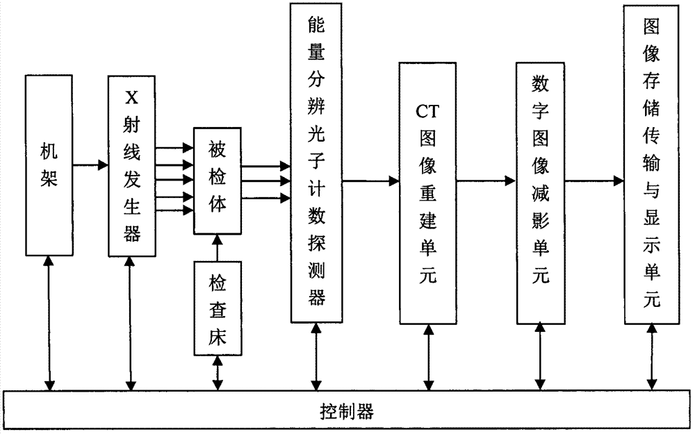
[0023] (3) Start the system under the control of the control unit, use the x-rays generated by the x-ray generator to quickly scan the subject, and the transmitted x-rays are accepted by the energy-resolving photon counting detector and counted in different energy regions; CT images The reconstruction unit collects the signal output by the energy-resolved photon counting detector to form two sets of transmission projection data in the high-energy area and low-energy area, and uses the analytical reconstruction algorithm or statistical reconstruction algorithm to reconstruct the tomographic images of the projection data in the high-energy area and low-energy ...


PUM


Abstract

The invention discloses a CT digital subtraction angiography (CT / DSA) imaging system and CT digital subtraction angiography (CT / DSA) imaging method. The CT / DSA imaging system is composed of an x-ray generator, an energy resolution photon counting detector, a CT image reconstruction unit, a digital image subtraction unit, an image storage, transmission and display unit, an examining bed, a machine frame and a controller, wherein an x-ray source generates continuous energy spectrum x rays for fast scanning the human body; the energy resolution photon counting detector is used for detecting the x photons penetrating through the human body and judging and counting the energy of the x photons; the CT image reconstruction unit is used for collecting signals output by the energy resolution photon counting detector, so that two groups of transmission projection data of a high-energy area and a low-energy area are formed, and the projection data of the high-energy area and the projection data of the low-energy area are subjected to tomography image reconstruction by adopting an analytical reconstruction algorithm or a statistical reconstruction algorithm; the digital image subtraction unit is used for applying different weights to the tomography reconstruction images of the high-energy area and the low-energy area of each same scanning position and carrying out subtraction calculation, and thus the CT digital subtraction angiography image is obtained.

Description

Technical field: [0001] The present invention relates to a medical X-ray imaging system and method, specifically to a CT digital subtraction angiography (computed tomography digital subtraction angiography, CT DSA) imaging system and method, which provides low-dose, high-quality imaging for the diagnosis and treatment of vascular lesions. effective means of imaging. Background technique: [0002] Vascular disease is one of the leading causes of death worldwide, and obtaining accurate patient vascular images is of great significance for clinical diagnosis and treatment. Angiographic imaging (DSA) is a large-scale real-time imaging diagnostic equipment, especially in coronary angiographic diagnosis, etc. It is an essential equipment, and it is also a platform for interventional therapy. It is mainly used for coronary stent implantation, minimally invasive valve implantation, interventional therapy for tumors, interventional therapy for cerebrovascular malformations and aneury...

Claims


Application Information

Patent Timeline
Patent Type & Authority Applications(China)
IPC IPC(8): A61B6/03A61B6/04
CPCA61B6/037A61B6/0407A61B6/40A61B6/4208A61B6/504A61B6/5211A61B6/5258A61B6/54
Inventor 周正东
Owner NANJING UNIV OF AERONAUTICS & ASTRONAUTICS
Why Eureka
Try Eureka