Tumor tissue 3D culture method and culture solution

A tumor tissue and culture method technology, applied in the field of biology, can solve the problems of easy floating of tissue samples, long period of species differences, poor tissue fixation, etc., to promote the process of research and development and precise treatment, easy operation, low cost effect

Active Publication Date: 2017-09-29
世翱(上海)生物医药科技有限公司
View PDF5 Cites 15 Cited by
  • Summary
  • Abstract
  • Description
  • Claims
  • Application Information

AI Technical Summary

Problems solved by technology

[0007] The technical problem mainly solved by the present invention is to provide a tumor tissue 3D culture method, which solves the problems of low accuracy, poor specificity, high cost, long cycle and species differences in the current conventional evaluation methods of anti-tumor drugs. To solve the problem of poor tissue fixation and easy floating of tissue samples using commonly used Matrigel glue, the culture carrier used in the present invention is hydrogel, which has good sample fixation, can also simplify steps, and is easy to operate and broaden the scope of application , can be widely used in the cultivation of various tumor tissues and drug efficacy evaluation

Method used


Image

  • Tumor tissue 3D culture method and culture solution
  • Tumor tissue 3D culture method and culture solution
  • Tumor tissue 3D culture method and culture solution

Examples


Embodiment 1

[0043] A method for 3D culture of intestinal cancer tissue is provided, comprising the steps of:

[0044] (1) Preparation of tissue culture plate

[0045] Take a 24-well plate, add 5-10 μL of phosphate buffer solution dropwise, put the high-temperature sterilized circular cover glass in the well plate, pat it to fit the bottom, and set it aside for later use;

[0046] (2) Preparation of tumor tissue blocks

[0047] Take intestinal cancer tumor tissue, wash it with PBS containing 1% double antibody and gentamicin, and cut it into 0.5-1 mm with ophthalmic surgical instruments 3 The tissue pieces were washed with normal saline, and the whole operation was performed on crushed ice.

[0048] (3) Tumor tissue block culture

[0049] The following is a comparative experiment of screening gels, and a comparative experiment is conducted for three commonly used gels for cell culture: Matrigel, hydrogel, and vitro-Gel 3D-RGD:

[0050] 3.1 Matrigel glue

[0051] Add 30-100 μL of undil...

Embodiment 2

[0063] A method for culturing various tumor tissues in 3D is provided, comprising the steps of:

[0064] (1) Preparation of tissue culture plate

[0065] Take a 24-well plate, add 5-10 μL of phosphate buffer solution dropwise, put the high-temperature sterilized circular cover glass in the well plate, pat it to fit the bottom, and set it aside for later use;

[0066] (2) Preparation of tumor tissue blocks

[0067] Take intestinal cancer, gastric cancer and esophageal cancer tumor tissues, wash them with PBS containing 1% double antibody and gentamycin, and cut them into 0.5-1 mm with ophthalmic surgical instruments 3 The tissue pieces were washed with normal saline, and the whole operation was performed on crushed ice.

[0068] (3) Tumor tissue block culture

[0069] Add 20-100 μL of hydrogel (diluted with 20% sterile sucrose solution 1:1-1:5) dropwise on the glass slide, carefully move the tumor tissue pieces of colon cancer, gastric cancer and esophagus cancer into the hy...

Embodiment 3

[0076] Example 3 Feasibility Analysis of 3D Cultured Tumor Tissues for Efficacy Evaluation of Antitumor Drugs

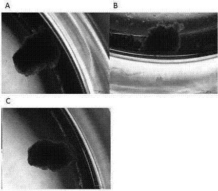
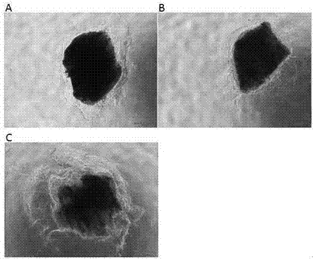
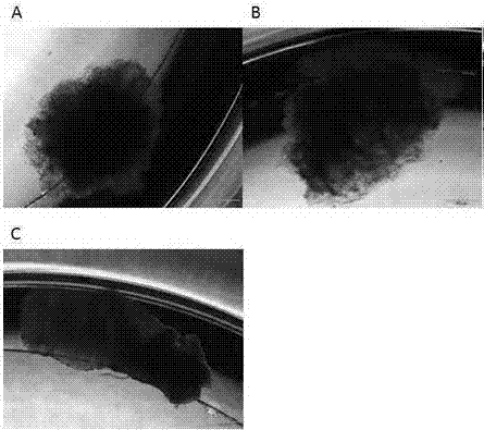
[0077] In Example 2, during the culture of intestinal cancer tissue, different concentrations and types of chemotherapeutic drugs were added to the culture medium, and the tissues were taken out at different times after culture, fixed in the above-mentioned fixative solution, and stained by HE staining, Ki67 immunohistochemical staining or Corresponding other staining, observe the tissue structure and cell morphology under the microscope, compare the morphological differences with the samples without anti-tumor drugs, evaluate the drug efficacy, count the number of proliferating cells, and calculate the cell proliferation rate, see Table 3, analyze the cell Feasibility of proliferation ratio as an evaluation method and index of antineoplastic drug potency. See for details Figure 13 , Figure 14 and Figure 15 .

[0078] table 3

[0079]

Number of K...


PUM


Abstract

The invention discloses a tumor tissue 3D culture method suitable for culturing various tumor tissues. The tumor tissue 3D culture method comprises the following steps: 1) dropwise adding hydrogel into a pore plate, transferring cut tumor tissue blocks into the hydrogel, dropwise adding a culture solution, and standing in a CO2 incubator in order to freeze the hydrogel; 2) adding the culture solution into the pore plate for culturing, taking the cultured tumor tissue blocks out and fixing, wherein the culture solution is prepared from an Advanced DMEM / F12 culture solution, 1*GlutaMAX, an epidermis growth factor (EGF), a fibroblast growth factor FGF10, N-acetyl-L-cysteine (NAC), prostaglandin E2 PGE2, Nicotinamide and 50*B27. Moreover, the invention further discloses a tumor tissue 3D culture solution. The method is suitable for a plurality of tumor tissues, can be used for rating the medicinal effects of antitumor drugs more rapidly and accurately, and overcomes the defects in cell 2D culturing and animal experiment rating methods; a more convenient and economical modeling method is provided for clinical medicament researches and new medicament development.

Description

technical field [0001] The present invention relates to the field of biology, in particular to a 3D tumor tissue culture method; in addition, the present invention also relates to a tumor tissue 3D culture solution. Background technique [0002] The World Cancer Report estimates that in 2012, the number of cancer cases in China was 3.065 million, accounting for about one-fifth of the global incidence; the number of cancer deaths was 2.205 million, accounting for about one-quarter of the global cancer deaths. Due to the aging population and other reasons, the current incidence and mortality of cancer in my country are showing a continuous increase trend. Currently, drug therapy accounts for 60% of cancer treatments and is still the main treatment option for tumors. However, in the existing standardized drug treatment, 70% of patients are ineffective, and in the process of cancer treatment, there is also a serious phenomenon of excessive chemotherapy. This excessive or unreas...

Claims


Application Information

Patent Timeline
Patent Type & Authority Applications(China)
IPC IPC(8): C12N5/09
CPCC12N5/0693C12N2500/32C12N2500/46C12N2501/02C12N2501/11C12N2501/119C12N2501/50C12N2513/00
Inventor 张皓雨徐霖康立清俞磊
Owner 世翱(上海)生物医药科技有限公司
Why Eureka
Try Eureka