
Microinjection device used for cell medicine precise injection and operation method of microinjection device

A microinjection and cell technology, applied in the field of cell biology research, can solve the problems of not specifying the method of making the injection needle, the difficult cells are transported in a straight line, and the cells cannot be moved flexibly, and achieves strong adjustability and high precision. , control flexible effects

Pending Publication Date: 2017-10-13
CHONGQING THREE GORGES MEDICAL COLLEGE
View PDF12 Cites 11 Cited by
  • Summary
  • Abstract
  • Description
  • Claims
  • Application Information

AI Technical Summary

Problems solved by technology

This cell microinjector uses all mechanical methods to control and inject cells. The existing problems are: the diameter of the micropipe transported by the cell transporter is fixed. For cells of different sizes, it is difficult to ensure that the cells will be arranged in a straight line in the transport pipeline. Front transport; cell capture and clamping adopts mechanical control, which cannot move cells flexibly. The method of mechanical control is very easy to damage cells. Mechanical control has the problem of being unable to stably clamp cells of different diameters. The designed clamping device can only be used for a certain The diameter of the cell clamping effect is the best; the patent does not explain the production method of the injection needle. For the microinjection of single cells, the control and precision of the injection should reach the level of soaring. It is very difficult to make high-precision ultra-fine needle tips; in addition, it is also difficult to pack cells tightly in the cell transporter in this patent

Method used


Image

  • Microinjection device used for cell medicine precise injection and operation method of microinjection device
  • Microinjection device used for cell medicine precise injection and operation method of microinjection device
  • Microinjection device used for cell medicine precise injection and operation method of microinjection device

Examples


Embodiment Construction

[0028] The preferred embodiments of the present invention will be described in detail below with reference to the accompanying drawings.

[0029] The reference signs in the accompanying drawings of the specification include:

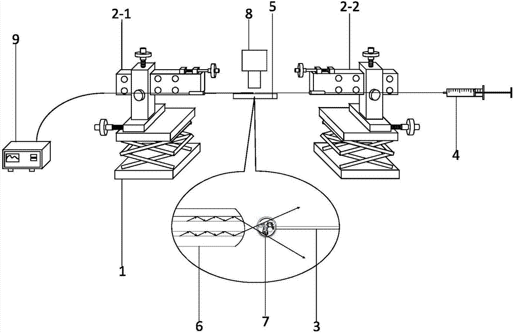
[0030] Lifting platform 1, three-dimensional high-precision translation platform 2, left translation platform 2-1, right translation platform 2-2, fiber optic microneedle 3, syringe 4, stage 5, optical tweezers 6, tumor cells 7, microscope 8, light source 9.

[0031] Such as figure 1 , figure 2As shown in the microinjection device for precise injection of cell drugs, the left lifting platform 1 and the right lifting platform 1 are both scissor-type folding experimental lifting platforms 1, the left lifting platform 1 is fixed by bolts to install the left fixed column, and the right lifting platform 1 The right fixed column is fixedly installed by bolts, and the middle parts of the left fixed column and the right fixed column are provided with grooves...


PUM


Abstract

The invention belongs to the technical field of cell biology study, and relates to a microinjection device used for cell medicine precise injection and an operation method of the microinjection device. A three-dimensional high-precision left displacement table is fixedly installed on a left lifting table, a three-dimensional high-precision right displacement table is fixedly installed on a right lifting table, a light source transmitting device is fixedly installed at the outer side of the left lifting table, optical tweezers are fixedly installed on the left lifting table, a syringe is arranged at the outer side of the right lifting table, an optical fiber microneedle is fixedly installed on the right lifting table, an objective table is installed between the left lifting table and the right lifting table, a microscope is installed above the objective table, during work, the displacement tables control the optical tweezers and the optical fiber microneedle, and therefore after the optical tweezers capture cells, precise injection of the optical fiber microneedle to the cells is achieved. The cell precise medicine injection device has the function of capturing and controlling the single cell and conducting precise medicine injection on the single cell, is high in precision and adjustability, and has a wide application prospect in the aspects of tumor cell precise medical experimental study and single cell microinjection medicine.

Description

technical field [0001] The invention belongs to the technical field of cell biology research, and relates to a microinjection device for precise injection of cell medicine and an operation method thereof. Background technique [0002] Tumor is one of the most important problems that endanger people's health. At present, 1 / 3 of the people in the world suffer from cancer, but 1 / 4 of them die from cancer. Tumor disease is easy to cause blockage and compression in its diseased area, which is caused by the excessive growth of tumor cells, which is similar whether it is a benign tumor or a malignant tumor. The common ones are blockage of esophagus, blockage of blood vessels and compression of nerves. It will affect the organ or tissue where the tumor disease is located in the adjacent organ, and the structure and function of the tumor cell will be destroyed. For example, liver cancer may experience systemic jaundice due to liver cell destruction and intrahepatic bile duct obstruc...

Claims


Application Information

Patent Timeline
IPC IPC(8): C12M1/00C12N15/89
CPCC12M1/00C12N15/89
Inventor 张永慧张竟原张楷金敦军魏勇刘春兰岳西刘奉
Owner CHONGQING THREE GORGES MEDICAL COLLEGE
Learn More