Marker used for in-vivo tracing and manual removal of CAR-T cells and application thereof

A tracer and cell technology, applied in the field of medical biology, to achieve the effect of reliable guarantee

Active Publication Date: 2017-10-24
SHANGHAI UNICAR THERAPY BIOPHARM TECH CO LTD
View PDF3 Cites 8 Cited by
  • Summary
  • Abstract
  • Description
  • Claims
  • Application Information

AI Technical Summary

Problems solved by technology

[0009] At present, there are no reports on CAR-T cell therapy that overcomes the above sho

Method used


Image

  • Marker used for in-vivo tracing and manual removal of CAR-T cells and application thereof
  • Marker used for in-vivo tracing and manual removal of CAR-T cells and application thereof
  • Marker used for in-vivo tracing and manual removal of CAR-T cells and application thereof

Examples


Embodiment 1

[0092] Example 1 Construction of CAR-T cells

[0093] 1. Construction, purification and detection methods of recombinant lentiviral vectors lvCARL1-lvCARL3.

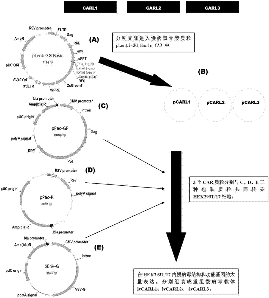
[0094] see image 3 , the construction method of the recombinant lentiviral vector of the present invention is as follows:

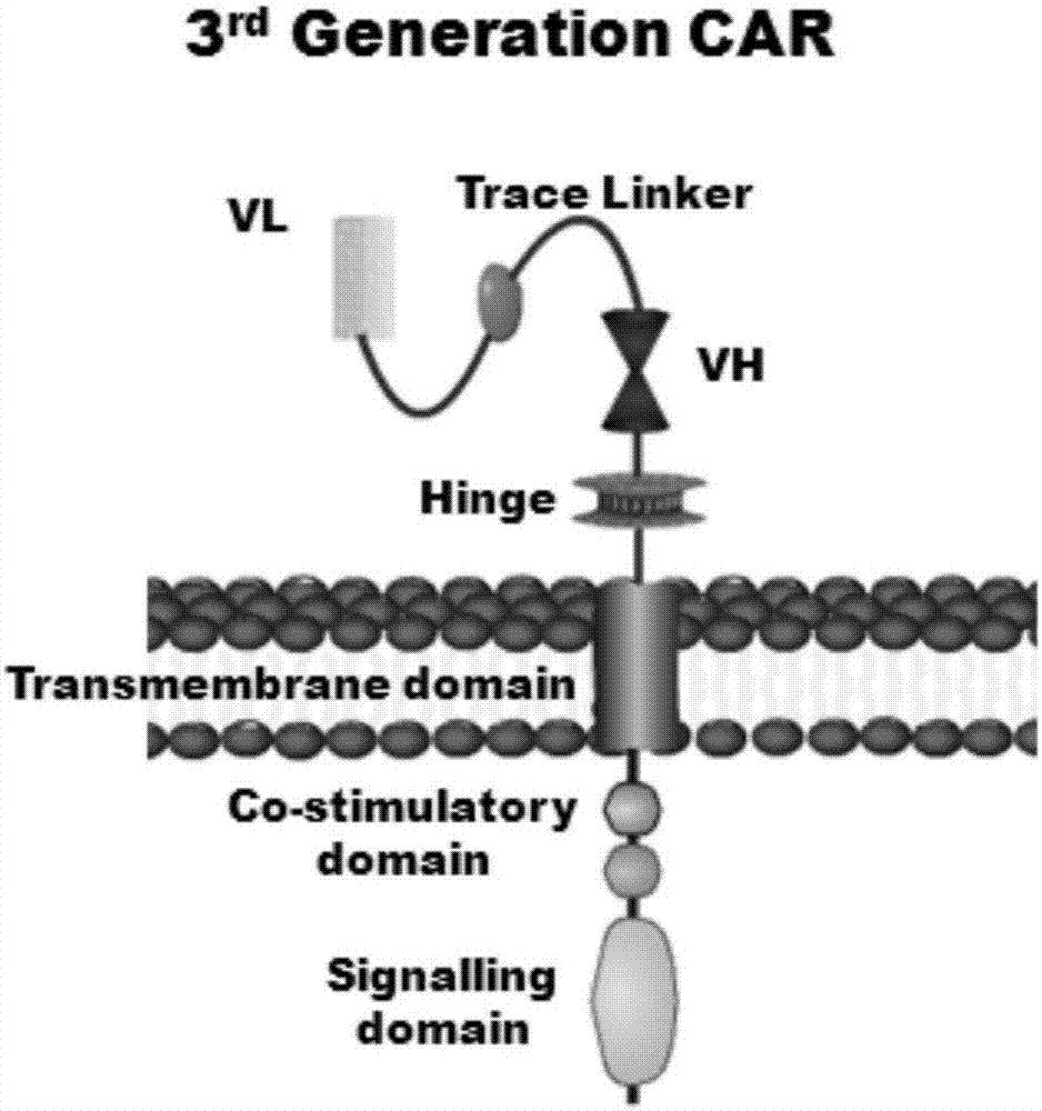
[0095] 1. Human EF1α promoter, CAR structure [CARL1, CARL2, CARL3] (CARL1, CARL2, CARL3 are the tracer hinge Trace-Linker1 containing the sequence shown in SEQ ID NO.18 respectively, as shown in SEQ ID NO.19 The traced hinge Trace-Linker2 of sequence shown, the CAR sequence of traced hinge Trace-Linker3 of sequence shown in SEQ ID NO.20, such as Figure 4 shown), cloned into lentiviral backbone plasmid pLenti-3G ​​basic, and obtained recombinant lentiviral plasmids pCARL1-pCARL3 respectively.

[0096] (1) The lentiviral backbone plasmid pLenti-3G ​​basic was double-digested with Cla I and EcoR I restriction endonucleases, and the product was subjected to 1.5% agarose gel electrophoresis to confi...

Embodiment 2

[0202] Pathogen detection and expression detection of CARL1-T~CARL3-T cells.

[0203] 1. Endotoxin detection;

[0204] (1), endotoxin working standard is 15EU / branch;

[0205] (2), Limulus reagent sensitivity λ=0.25EU / ml, 0.5ml / tube

[0206] (3) Dilution of endotoxin standard substance: Take one endotoxin standard substance, dilute it with BET water in proportion to dissolve into 4λ and 2λ respectively, seal with parafilm, shake and dissolve for 15min; each step of dilution should be mixed in the vortex Mix on the mixer for 30s;

[0207] (4) Adding samples: Take several LAL reagents, add 0.5 ml of BET water to each tube to dissolve, and distribute to several endotoxin-free test tubes, each tube has 0.1 ml. Two of them are negative control tubes, add 0.1ml of BET water;

[0208] Two are positive control tubes, add 0.1ml of endotoxin working standard solution with 2λ concentration;

[0209] 2 tubes are sample positive control tubes, add 0.1ml sample solution containing 2λ e...

Embodiment 3

[0239] Example 3 Functional detection of CARL1-T~CARL3-T cells.

[0240] 1. Evaluation of target cell killing effect.

[0241] (1) Culture target cells separately [CD117 + K562, K562 cells] and effector cells [CARL1-T~CARL3-T cells];

[0242] (2) Collect target cells 4x10 5 cells and CARL1-T~CARL3-T cells 2.8x10 6 cells, 800g, centrifuge for 6min, discard the supernatant;

[0243] (3) Resuspend the target cells and effector cells in 1ml D-PBS(-) solution, centrifuge at 800g for 6min, discard the supernatant;

[0244] (4) Repeat step 3 once;

[0245] (5) Resuspend effector cells with 700ul medium (AIM-V medium + 1-10% FBS), and resuspend target cells with 2ml medium (AIM-V medium + 1-10% FBS);

[0246] (6) Set up experimental wells with effect-to-target ratios of 1:1, 5:1, and 10:1, and set up a control group (K562 cells), with 3 replicate wells in each group;

[0247] (7) 250g, 5min plate centrifugation;

[0248] (8) Cultivate for 4 hours in a 5% CO2 incubator at 37°C;...


PUM

PropertyMeasurementUnit
Sensitivityaaaaaaaaaa
Absorbanceaaaaaaaaaa

Abstract

The invention discloses a marker used for in-vivo tracing and manual removal of CAR-T cells. The marker comprises a trace-linker 1 with a sequence as shown in SEQ ID No. 18, a trace-linker 2 with a sequence as shown in SEQ ID No. 19 and a trace-linker 3 with a sequence as shown in SEQ ID No. 19. The invention also discloses a CAR-T therapy vector including the marker and a construction method thereof. Moreover, the invention further discloses application of the marker to preparation of the CAR-T therapy vector and drugs used for treating triple-negative breast cancer. The marker provided by the invention can realize controllable in-vivo tracing, in-vitro separation and manual removal of CAR-T cells without influence on the tumor killing effect of the CAR-T cells, so the security of CAR-T cell therapy is greatly improved, and a powerful pool can be provided for deeper analysis of the process of CAR-T cell therapy. The vector provided by the invention can express a targeting chimeric antigen receptor of CD117 in human T lymphocytes, guides and activates killing effect of T lymphocytes on CD117 positive cells, and is applicable to treatment of triple-negative breast cancer in clinical practice.

Description

technical field [0001] The invention belongs to the field of medical biology, and in particular relates to a biological label, in particular to a label for tracing and artificially clearing CAR-T cells in vivo. Furthermore, the invention also relates to the application of the label. Background technique [0002] The theoretical basis of tumor immunotherapy is that the immune system has the ability to recognize tumor-associated antigens and regulate the body's ability to attack tumor cells (highly specific cytolysis). In the 1950s, Burnet and Thomas put forward the theory of "immune surveillance", thinking that the mutated tumor cells that often appear in the body can be recognized and eliminated by the immune system, which laid the theoretical foundation for tumor immunotherapy [Burnet FM. Immunological aspects of malignant disease. Lancet, 1967; 1:1171-4]. Subsequently, various tumor immunotherapies, including cytokine therapy, monoclonal antibody therapy, adoptive immuno...

Claims


Application Information

Patent Timeline
IPC IPC(8): C12N15/13C12N15/867C12N15/66A61K35/17A61P35/00
CPCA61K35/17C07K16/2887C12N15/66C12N15/86C12N2740/15043
Inventor 祁伟俞磊康立清林高武余宙
Owner SHANGHAI UNICAR THERAPY BIOPHARM TECH CO LTD
Why Eureka
Try Eureka