Time-resolved fluoroimmunoassay method for detecting avian influenza virus H7N9

A technology of time-resolved fluorescence and avian influenza virus, which is applied in the direction of antiviral immunoglobulin, analytical materials, immunoglobulin, etc., can solve the problem of lack of detection kits for H7N9, reduce the risk of H7N9 infection, be easy to promote, The effect of reducing economic loss

Active Publication Date: 2017-11-14
GUANGZHOU YOUDI BIOTECH CO LTD
View PDF6 Cites 8 Cited by
  • Summary
  • Abstract
  • Description
  • Claims
  • Application Information

AI Technical Summary

Problems solved by technology

[0007] However, there are currently no kits and methods for detecting H7N9 using TRFIA in the market

Method used


Image

  • Time-resolved fluoroimmunoassay method for detecting avian influenza virus H7N9
  • Time-resolved fluoroimmunoassay method for detecting avian influenza virus H7N9
  • Time-resolved fluoroimmunoassay method for detecting avian influenza virus H7N9

Examples


Embodiment 1

[0038] Example 1 Construction of single-chain antibody phage library of highly pathogenic avian influenza virus H7N9, screening of single-chain antibody and induced expression

[0039] (1) Construction of anti-human avian influenza virus H7N9 single-chain antibody phage library

[0040] ① Isolation of total RNA: Human total mRNA was isolated from 10 mL of whole blood of a patient who had recovered from avian influenza H7N9 using a blood RNA extraction kit (Qiagen Corp).

[0041] ② Synthesize the first cDNA strands of IgG light and heavy chain genes of anti-human avian influenza virus H7N9 antibody: the RNA extracted in the above step is used as a template, and in order to increase the specificity of amplification, reverse transcription primers (HomoIgG1-4H1F, HomoCk-F and HomoCλ-F) to specifically amplify the first strand of cDNA of human IgG1 constant region and light chain k, lambda constant region.

[0042] The above reverse transcription primer sequence is:

[0043] Homo...

Embodiment 2

[0075] Example 2 Time-resolved fluorescent immunoassay kit for highly pathogenic avian influenza virus H7N9

[0076]The time-resolved fluorescent immunoassay kit for H7N9 contains the following components:

[0077] (1) Anti-H7N9 single-chain antibody

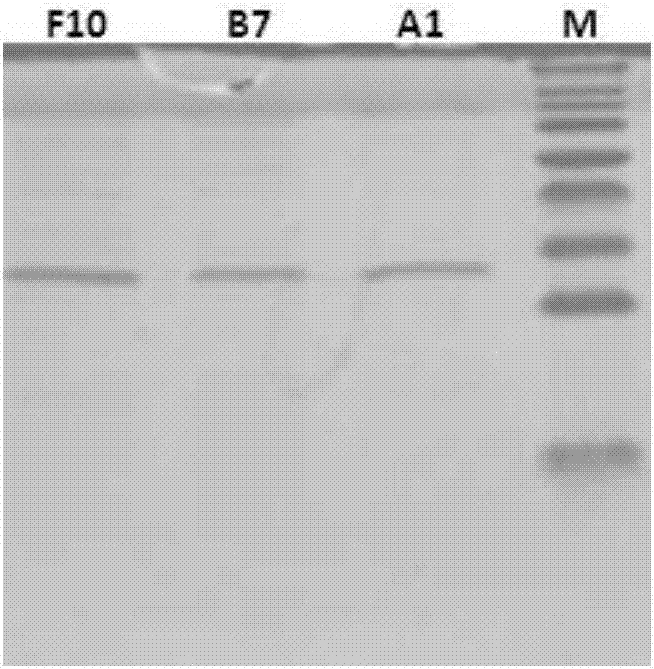
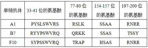
[0078] The anti-H7N9 single-chain antibody was selected from one of the single-chain antibodies A1, B7, and F10 described in Experimental Example 1 above.

[0079] (2) Enzyme-linked plates coated with antibodies

[0080] Use 50mmol / L, pH 9.6 carbonate buffer to dilute the coating antibody B7 (A1 or F10 can also be used) to 1-5μg / ml, then add 100μl / well to each well of the Elisa plate, 4 overnight at ℃, discard the coating solution, add blocking solution, 200 μl / well, block overnight, discard the coating solution, wash and pat dry, and store in vacuum at -20℃.

[0081] (3) Eu 3+ labeled antibody

[0082] Add 0.5 mg of the labeled antibody into a centrifuge tube with a filter membrane from Millipore, and centrifuge at 8000 r / ...

Embodiment 3

[0091] Example 3 Time-resolved fluorescent immunoassay method for highly pathogenic avian influenza virus H7N9

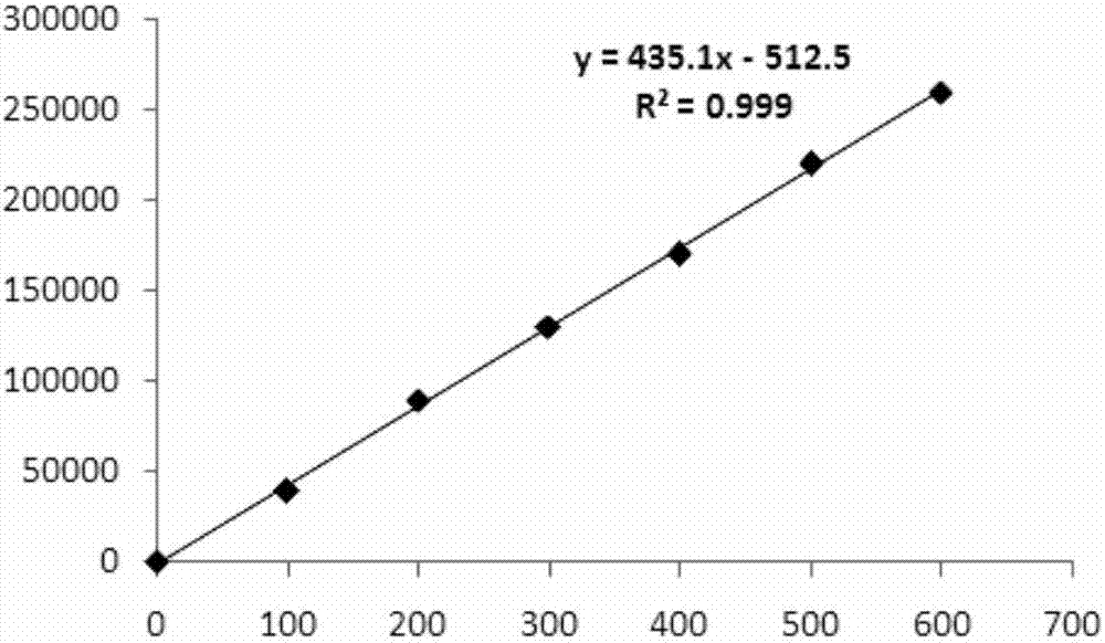
[0092] The operation steps of the time-resolved fluorescent immunoassay kit for quantitative detection of H7N9 virus of the present invention are as follows: add 25 μl of reference standard or sample to be tested to the coated enzyme-linked plate, then add 200 μl of analysis buffer, incubate with shaking for 1 hour, wash with Wash 4 times, use the analysis buffer to dilute the Eu 3+ Label the antibody to 1-5 μg / ml, add 200 μl / well, incubate with shaking for 1 h, wash with washing solution 6 times, and finally add 200 μl / well of enhancement solution (Perkin Elmer Company) and shake for 5 minutes before detecting on a time-resolved fluorescence detector.

[0093] The effect detection of the kits prepared in the above examples is performed below.


PUM

PropertyMeasurementUnit
Sensitivityaaaaaaaaaa
recovery rateaaaaaaaaaa

Abstract

The invention discloses a time-resolved fluoroimmunoassay method for detecting avian influenza virus H7N9. The amino acid sequence of a single-chain antibody for detecting the avian influenza virus H7N9 is shown as SEQ ID NO:1, SEQ ID NO:2 or SEQ ID NO:3. The method comprises the following steps: adding a H7N9 virus standard or a to-be-detected sample to an assay plate coated with the single-chain antibody; then adding an analysis buffer solution and shaking at room temperature for reaction; after washing with a washing solution; adding 100-200mu l / hole Eu<3+> labeled antibody diluted by the analysis buffer solution according to a volume ratio of 1:(50 to 100); after shaking at the room temperature for reaction, and washing with the washing solution; finally, adding an enhancement solution for fluorescence detection. The method and kit disclosed by the invention have the advantages of high sensitivity, high speed, convenience and easiness in promotion, and can detect the H7N9 virus in the samples of human, poultry, animals and external environment in time.

Description

technical field [0001] The invention belongs to the field of biotechnology, and in particular relates to a time-resolved fluorescent immunoassay method for detecting avian influenza virus H7N9. Background technique [0002] Influenza A H7N9 virus is a new type of avian influenza virus first discovered in Shanghai and Anhui in eastern China in March 2013. Infection with this virus will cause pneumonia, respiratory failure, acute respiratory distress syndrome (Acute Respiratory Distress Syndrome, ARDS) ). As of July 2014, the World Health Organization (WHO) announced 450 cases of human infection with H7N9 avian influenza and 165 deaths. H7N9 is a subtype of avian influenza virus, which mainly occurs in birds and can also occur in mammals. H7N9 has higher infectivity to humans, and it is mainly sporadic cases in humans. Human beings are infected by contact with infected diseases, dead poultry excrement, secretions, or the environment or objects contaminated by their excrement...

Claims


Application Information

Patent Timeline
Patent Type & Authority Applications(China)
IPC IPC(8): C07K16/10G01N33/569
CPCC07K16/1018C07K2317/622
Inventor 刘晓柱梁焕坤李康燕陈翠翠李来庆王珊霞钟树海程承武刘细潘
Owner GUANGZHOU YOUDI BIOTECH CO LTD
Why Eureka
Try Eureka