Digital brain visualization method, device, computing device and storage medium

A brain visualization and digital technology, applied in the field of medical image processing, can solve the problems of low accuracy of fusion of brain tissue and cerebral blood vessels, poor digital brain visualization effect, etc., and achieve the effect of improving the effect and improving the accuracy.

Active Publication Date: 2020-08-18
SHENZHEN INST OF ADVANCED TECH CHINESE ACAD OF SCI
View PDF5 Cites 0 Cited by
  • Summary
  • Abstract
  • Description
  • Claims
  • Application Information

AI Technical Summary

Problems solved by technology

[0005] The purpose of the present invention is to provide a digital brain visualization method, device, computing equipment and storage medium, aiming to solve the problem of poor digital brain visualization effect due to the low accuracy of brain tissue and cerebrovascular fusion in the existing digital brain visualization technology The problem

Method used


Image

  • Digital brain visualization method, device, computing device and storage medium
  • Digital brain visualization method, device, computing device and storage medium
  • Digital brain visualization method, device, computing device and storage medium

Examples


Embodiment 1

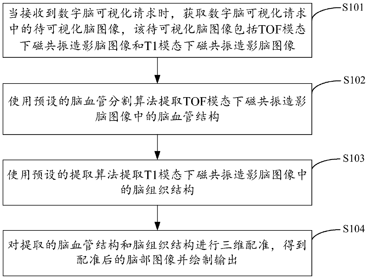
[0026] figure 1 The implementation process of the digital brain visualization method provided by Embodiment 1 of the present invention is shown. For the convenience of description, only the parts related to the embodiment of the present invention are shown, and the details are as follows:

[0027] In step S101, when a digital brain visualization request is received, the brain image to be visualized in the digital brain visualization request is obtained, and the brain image to be visualized includes the MRI brain image under the TOF mode and the MRI brain image under the T1 mode. image.

[0028] The embodiments of the present invention are suitable for medical image processing systems, especially for digital brain visualization systems, so as to facilitate the visualization of magnetic resonance imaging brain images. In the embodiment of the present invention, when digital brain visualization is required, the brain images to be visualized input into the system include MRI brai...

Embodiment 2

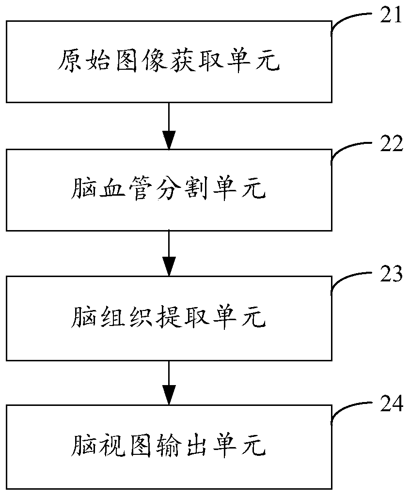
[0040] figure 2 The structure of the digital brain visualization device provided by Embodiment 2 of the present invention is shown. For the convenience of description, only the parts related to the embodiment of the present invention are shown, including:

[0041] The original image acquisition unit 21 is used to acquire the brain image to be visualized in the digital brain visualization request when receiving the digital brain visualization request, the brain image to be visualized includes the MRI brain image under the TOF mode and the MRI brain image under the T1 mode. Resonance-enhanced brain images.

[0042] In the embodiment of the present invention, when digital brain visualization is required, the brain images to be visualized acquired by the original image acquisition unit 21 include MRI brain images in TOF mode and MRI brain images in T1 mode, In order to facilitate subsequent extraction of cerebrovascular structures and brain tissue structures. The MRI image unde...

Embodiment 3

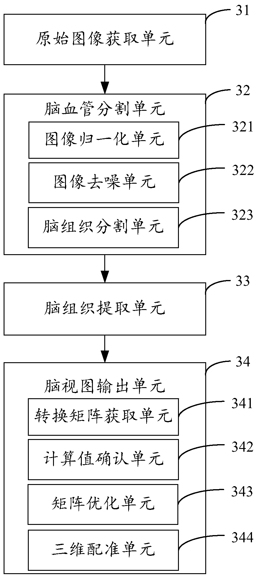
[0050] image 3 The structure of the digital brain visualization device provided by the third embodiment of the present invention is shown. For the convenience of description, only the parts related to the embodiment of the present invention are shown, including:

[0051] The original image acquisition unit 31 is used to acquire the brain image to be visualized in the digital brain visualization request when receiving the digital brain visualization request, the brain image to be visualized includes the MRI brain image under the TOF mode and the MRI brain image under the T1 mode. Resonance-enhanced brain images.

[0052] In the embodiment of the present invention, when digital brain visualization is required, the brain images to be visualized acquired by the original image acquisition unit 31 include MRI brain images in TOF mode and MRI brain images in T1 mode, In order to facilitate subsequent extraction of cerebrovascular structures and brain tissue structures. The MRI ima...


PUM


Abstract

The invention is suitable for the technical field of medical image processing and provides a digital brain visualization method and device, a computing device and a storage medium. The method comprises the following steps: when receiving a digital brain visualization request, obtaining a brain image to be visualized in the digital brain visualization request, wherein the brain image to be visualized comprises a magnetic resonance imaging brain image under a TOF modal and a magnetic resonance imaging brain image under a T1 modal; extracting a brain blood vessel structure in the magnetic resonance imaging brain image under the TOF modal through a preset brain blood vessel segmentation algorithm, and extracting a brain tissue structure in the magnetic resonance imaging brain image under the T1 modal through a preset extraction algorithm; and carrying out three-dimensional registration on the extracted brain blood vessel structure and the brain tissue structure to obtain a brain image after registration, and carrying out image drawing and output, thereby improving brain tissue and brain blood vessel registration and fusion accuracy, improving digital brain visualization accuracy and improving the digital brain visualization effect.

Description

technical field [0001] The invention belongs to the technical field of medical image processing, and in particular relates to a digital brain visualization method, device, computing equipment and storage medium. Background technique [0002] With the development of medical science and technology and the continuous updating of diagnosis and treatment equipment, modern surgery is developing towards minimally invasive, microscopic and replacement. In addition to the development of surgical guidance systems, virtual surgery and robotic surgery, familiarity with the topology of blood vessels and the entire tissue space can greatly improve the success rate of surgery. [0003] At present, in terms of blood vessel visualization research, the relatively breakthrough is the blood vessel marking technology first reported in 2002, which successfully realized the "Chinese digital human", combined with anatomical knowledge, and finally realized the digital human by slicing the real human...

Claims


Application Information

Patent Timeline
Patent Type & Authority Patents(China)
IPC IPC(8): G06T7/143G06T7/33G06T15/00
CPCG06T7/143G06T7/337G06T15/005G06T2207/10088G06T2207/20221G06T2207/30016G06T2207/30101
Inventor 王澄李迟迟李江宁周寿军
Owner SHENZHEN INST OF ADVANCED TECH CHINESE ACAD OF SCI
Why Eureka
Try Eureka