Biological colorimetric sensor, preparation method and application in glucose detection

A colorimetric sensor, glucose technology, applied in the measurement of color/spectral characteristics, analysis by making materials undergo chemical reactions, material analysis by observing the impact on chemical indicators, etc., can solve the limitations of practical applicability, lengthy detection process, high detection limit and other issues, to achieve the effect of low cost, high sensitivity and low detection limit

Inactive Publication Date: 2018-01-09
ANHUI NORMAL UNIV
View PDF4 Cites 2 Cited by
  • Summary
  • Abstract
  • Description
  • Claims
  • Application Information

AI Technical Summary

Problems solved by technology

However, most of the existing glucose sensors involve complex and time-consuming detection processes, and most of them have disadvantages such as low selectivity and relatively high detection limits, which limit their practical applicability.

Method used


Image

  • Biological colorimetric sensor, preparation method and application in glucose detection
  • Biological colorimetric sensor, preparation method and application in glucose detection
  • Biological colorimetric sensor, preparation method and application in glucose detection

Examples


Embodiment 1

[0038] A method for preparing a biological colorimetric sensor, comprising the following steps:

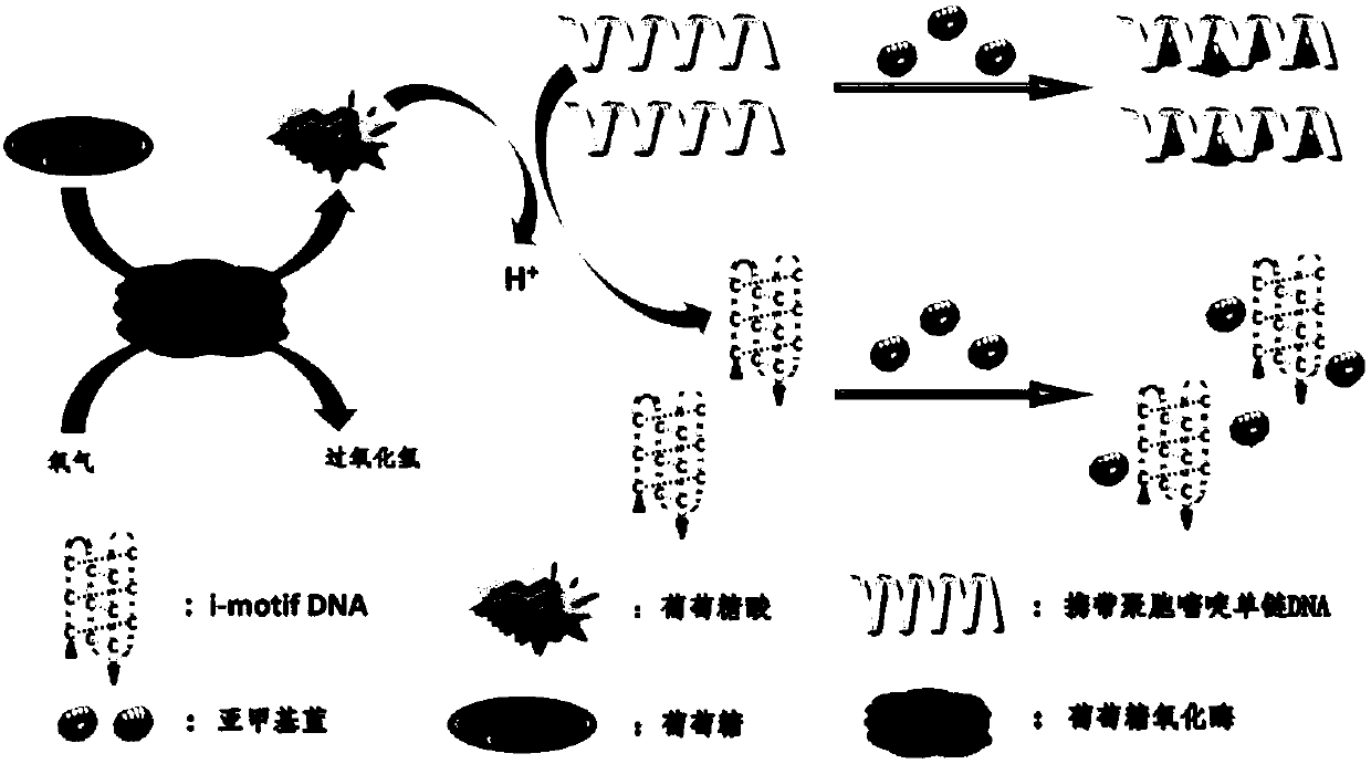
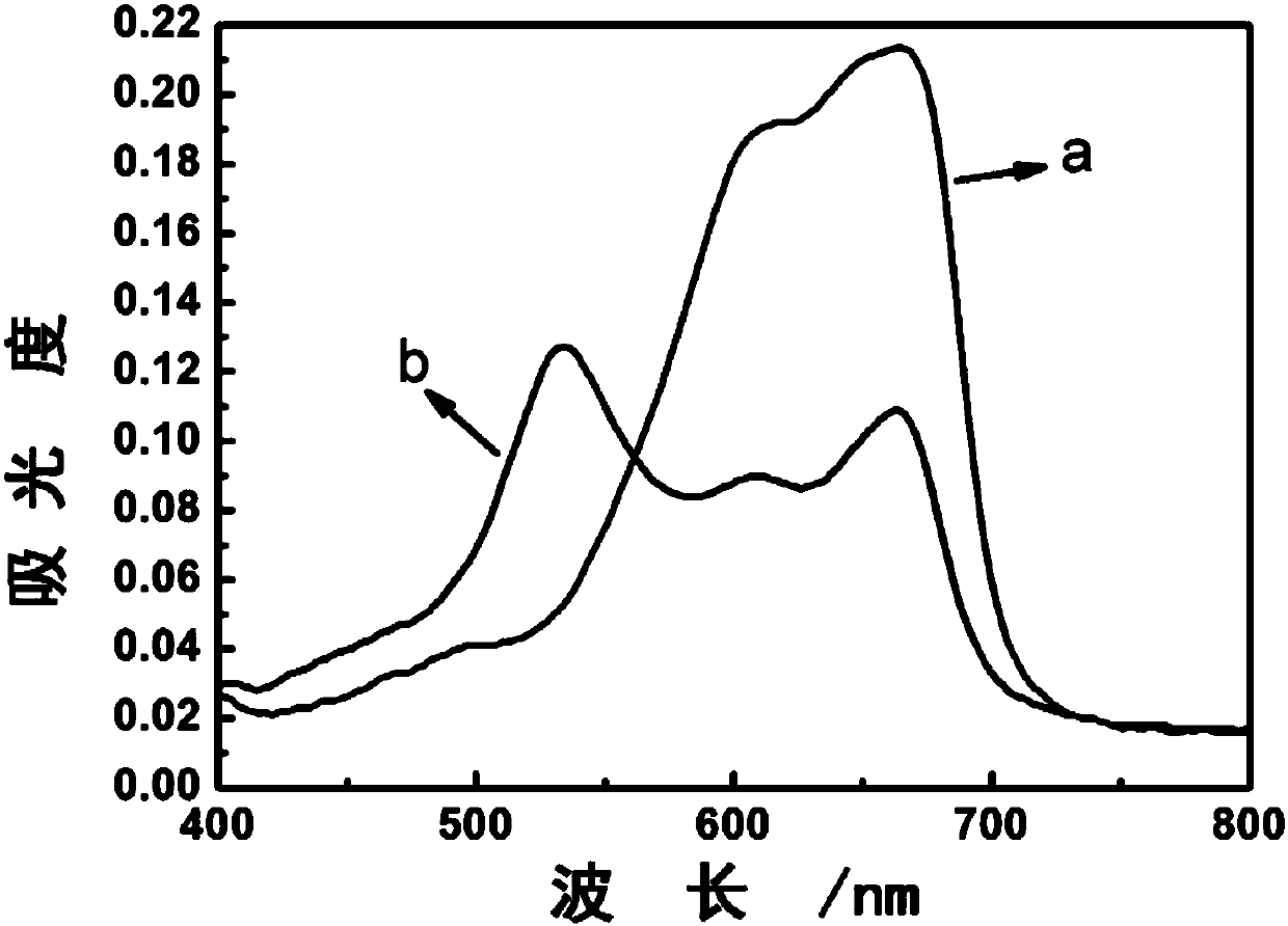
[0039] Take 50μL 20mM glucose solution and add it to 78μL Tris-EDTA (10mM Tris, 1mM EDTA, pH 8.0) buffer solution, then add 20μL 3mM glucose oxidase solution, incubate at 25°C for 30min, then add 50μL 100μM methylene blue solution, mix well, and then add 2 μL of 100 μM ssDNA buffer solution to obtain a mixed solution, observe the color of the solution, and use a biocolorimetric sensor to detect the ultraviolet-visible absorption spectrum.

[0040] Due to the addition of a mixture of glucose and glucose oxidase, the gluconic acid produced by the reaction lowers the pH of the solution and induces ssDNA to fold into a tight four-overlapping structure called "i-motif" DNA, which will hinder ssDNA and methylene blue to varying degrees interaction, the solution color changed from purple to blue. In the UV-Vis absorption spectrum, the typical absorption peak of methylene blue reappeared...

Embodiment 2

[0042] The pH of the control buffer solution is different, and other steps are the same as in Example 1, and obtain the corresponding ultraviolet-visible absorption spectra of different pH values; see Figure 3A , Figure 3B .

Embodiment 3

[0044] An application of a biological colorimetric sensor for detecting glucose, an application for detecting glucose.

[0045] The specific detection method is:

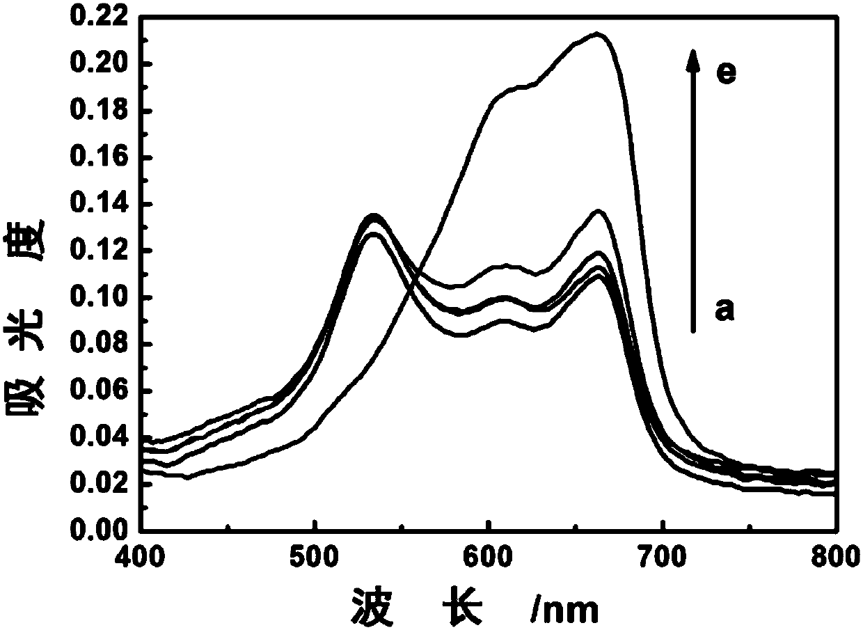
[0046] 1) Glucose was dissolved in deionized water to prepare 0.05, 0.1, 1, 2.5, 3, 4, 5, 8, 10 and 20 mM glucose solutions.

[0047] 2) Add 20 μL of the above-prepared concentration gradient glucose solution to 108 μL Tris-EDTA (10 mM Tris, 1 mM EDTA, pH 8.0) buffer solution, and then add 20 μL of 3 mM glucose oxidase solution, and incubate at 25°C for 30 min;

[0048] 3) Add 50 μL of 100 μM methylene blue solution, mix well, and then add 2 μL of 100 μM ssDNA buffer solution to obtain a mixed solution, observe the color of the solution, and detect the ultraviolet-visible absorption spectrum. Specific results such as Figure 4A .

[0049] The gluconic acid generated by the reaction of glucose and glucose oxidase reduces the pH value of the solution and induces ssDNA to fold into an "i-motif" structure, which will...


PUM


Abstract

The invention discloses a biological colorimetric sensor, a preparation method and application in glucose detection. The preparation method comprises the following steps: putting a mixture of a glucose solution and a glucose oxidase solution into a buffer solution, performing culture, adding a methylene blue solution, uniformly mixing, and further adding an ssDNA (Single-Stranded DeoxyribonucleicAcid) buffer solution, thereby obtaining a mixed solution, namely the biological colorimetric sensor. Compared with the prior art, the biological colorimetric sensor preparation method disclosed by the invention is low in cost and simple to design, the colors of solutions can be changed with glucose of different concentrations, and the change can be read with naked eyes. Under interaction of methylene blue and single-chain DNA, and by virtue of conformation interconversion of the single-chain DNA with polycytosine, a sensor for detecting glucose can be prepared. Results show that the sensor iscapable of sensitively detecting glucose with the concentration of 5mu M to 0.8mM, and also is simple to operate, high in sensitivity and lo win detection limit.

Description

technical field [0001] The invention belongs to the technical field of biosensor preparation, and specifically relates to a biocolorimetric sensor, a preparation method and its application for detecting glucose, which is based on the interaction between methylene blue and single-stranded DNA and utilizes the conformation of single-stranded DNA carrying polycytosine The constructed glucose sensor was converted and able to detect glucose. Background technique [0002] Glucose, as a ubiquitous fuel in the metabolic pathways of organisms, is the basic need to meet life activities. An unusual fluid level of glucose is a warning sign for human health. For example, in clinical medicine, diabetes seriously endangers human health, and its diagnosis and treatment have always been a major problem in the medical field. Diabetes can be effectively monitored and treated by accurately measuring the blood sugar content of diabetic patients. It can be seen that blood glucose monitoring is...

Claims


Application Information

Patent Timeline
Patent Type & Authority Applications(China)
IPC IPC(8): G01N21/78G01N21/31
Inventor 王广凤王琴
Owner ANHUI NORMAL UNIV
Why Eureka
Try Eureka