Magnetic resonance imaging method, device and system

A magnetic resonance imaging and magnetic resonance signal technology, which is applied in measuring devices, measuring magnetic variables, medical science, etc., can solve the problems of amplifying image noise, enlarging errors, reducing image quality, etc., achieving calibration data optimization, improving quality, and improving The effect of precision

Active Publication Date: 2018-02-02
SHANGHAI UNITED IMAGING HEALTHCARE
View PDF9 Cites 12 Cited by
  • Summary
  • Abstract
  • Description
  • Claims
  • Application Information

AI Technical Summary

Problems solved by technology

Among them, vascular pulsation will cause pulsation artifacts in the calibration coefficient itself, which will affect the accuracy of the calibration data. In the subsequent operations based on the calibration coefficient, the error will be enlarged, the image noise will be amplified, and the image quality will be reduced.

Method used


Image

  • Magnetic resonance imaging method, device and system
  • Magnetic resonance imaging method, device and system
  • Magnetic resonance imaging method, device and system

Examples


Embodiment 1

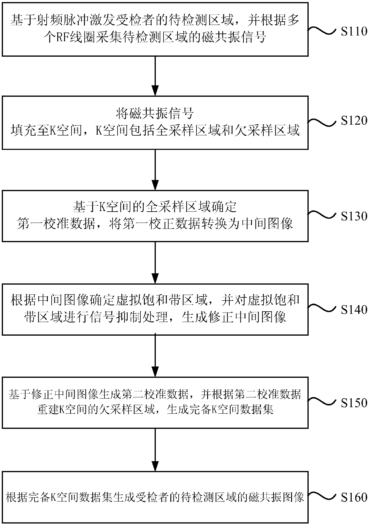
[0055] figure 2It is a flow chart of a magnetic resonance imaging method provided in Embodiment 1 of the present invention. The method can be executed by a magnetic resonance imaging device provided in the embodiment of the present invention, and the device can be realized by software and / or hardware. see figure 2 , the method specifically includes:

[0056] S110. Excite the subject's region to be detected based on radio frequency pulses, and collect magnetic resonance signals of the region to be detected according to a plurality of RF coils.

[0057] Wherein, a magnetic resonance scan is performed on the subject through a magnetic resonance imaging system, and the magnetic resonance imaging system includes a magnetic resonance scanning device and an image generating device. The magnetic resonance scanning device includes but is not limited to a control unit, a radio frequency transmitting unit, and a magnetic resonance signal acquisition unit. When the examinee moves to ...

Embodiment 2

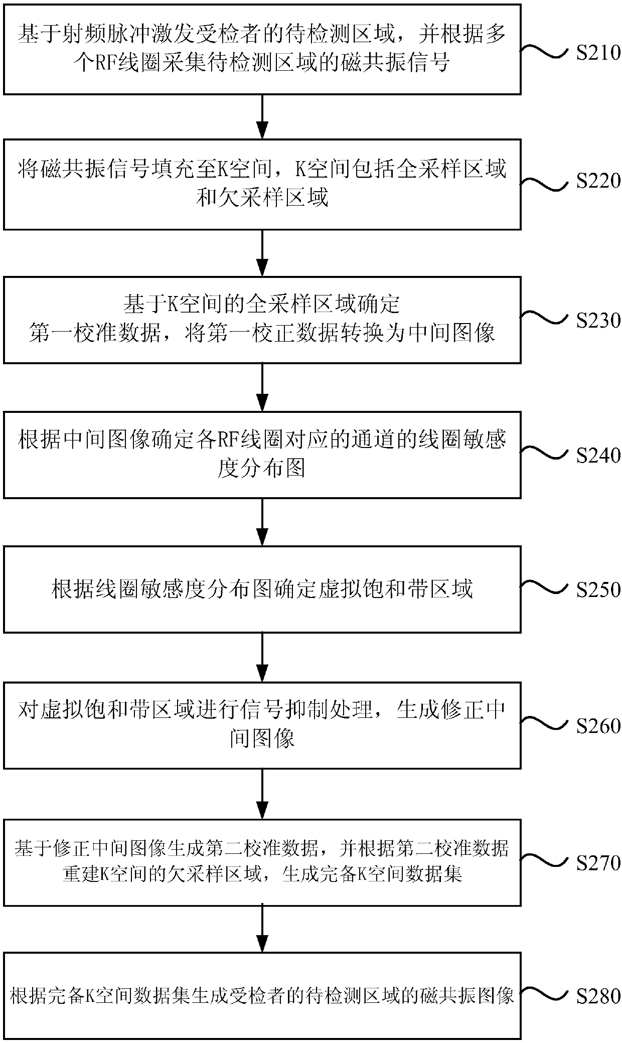
[0081] image 3 It is a flow chart of a magnetic resonance imaging method provided in Embodiment 2 of the present invention. On the basis of the above embodiments, a method for determining a virtual saturation band area according to an intermediate image is further provided. Accordingly, the method specifically includes:

[0082] S210. Excite the subject's region to be detected based on radio frequency pulses, and acquire magnetic resonance signals of the region to be detected according to a plurality of RF coils.

[0083] S220. Perform phase encoding on the magnetic resonance information, acquire multiple phase data lines, and fill the phase data lines into a K space, where the K space includes a full sampling area and an under sampling area.

[0084] S230. Determine first calibration data based on the full sampling area of ​​the K-space, and convert the first calibration data into an intermediate image. In this embodiment, zero-fill processing is not performed on the high-...

Embodiment 3

[0113] Figure 4 It is a flow chart of a magnetic resonance imaging method provided in the third embodiment of the present invention. The method can be executed by a magnetic resonance imaging system provided in the embodiment of the present invention. The magnetic resonance imaging system can use software and / or hardware accomplish. see Figure 4 , the method specifically includes:

[0114] S310. Exciting precession nuclear spins in the region to be detected of the subject.

[0115] S320. Use multiple RF coils to simultaneously acquire multiple response signals, where the multiple response signals represent magnetic resonance signals generated by precessing nuclear spins in the region to be detected.

[0116] S330. Fill the multiple response signals into K-space, and acquire an undersampled K-space data set.

[0117] S340. Acquire calibration data for each under-sampled data set.

[0118] Among them, the undersampling data and the calibration data are obtained separately...


PUM


Abstract

The embodiment of the invention discloses a magnetic resonance imaging method, device and system. The magnetic resonance imaging method comprises the steps of exciting a region to be detected of a subject based on radio frequency pulse, and collecting magnetic resonance signals of the region to be detected according to a plurality of RF coils; filling the magnetic resonance signals into a K-space,wherein the K-space comprises an all-sampling region and an under-sampling region; determining first calibration data based on the all-sampling region of the K-space, and converting the first calibration data into an intermediate image; determining a virtual saturation zone according to the intermediate image, and performing signal suppression processing on the virtual saturation zone to generatea modified intermediate image; generating second calibration data based on the modified intermediate image, reconstructing an under-sampling region of the K-space according to the second calibrationdata, and generating a complete K-space data set; and generating a magnetic resonance image of the region to be detected according to the complete K-space data set. The quality of the magnetic resonance image is improved according to the embodiment of the invention.

Description

technical field [0001] Embodiments of the present invention relate to magnetic resonance imaging technology, and in particular to a magnetic resonance imaging method, device and system. Background technique [0002] Magnetic resonance imaging (Magnetic Resonance Imaging, MRI) is a technology that uses magnetic resonance phenomena for imaging. It has the characteristics of non-invasiveness, no ionization, and radiation. Advantages, especially for the soft tissue level display is very good, can greatly improve the diagnostic efficiency, often used in clinical diagnosis. Magnetic resonance imaging is usually affected by factors such as respiration, vascular pulsation, or involuntary movement. For example, vascular pulsation generally appears to produce pulsation artifacts along the phase encoding direction. If the basic acquisition and reconstruction technology is applied during the MRI process, technicians or doctors with rich clinical experience can identify pulsation artifa...

Claims


Application Information

Patent Timeline
Patent Type & Authority Applications(China)
IPC IPC(8): G01R33/565A61B5/055
CPCA61B5/055G01R33/56509
Inventor 翟人宽
Owner SHANGHAI UNITED IMAGING HEALTHCARE
Why Eureka
Try Eureka