MnO2 wrapped polydopamine nano-particle, preparation method and application
A technology of polydopamine and nanoparticles, which is applied in the field of functional nanomaterials and its preparation, can solve the problems of poor biocompatibility of inorganic materials, restrictions on the application of photothermal preparations, and the enrichment of organic photosensitive molecules with short blood half-lives. Effects of biocompatibility and photothermal performance
- Summary
- Abstract
- Description
- Claims
- Application Information
AI Technical Summary
Problems solved by technology
Method used
Image
Examples
Embodiment 1
[0025] Preparation of polydopamine (PDA) nanoparticles:
[0026] (1) Dissolve 180 mg of dopamine hydrochloride in 90 mL of water, bathe in water at 50°C for 20 minutes, quickly add 760 μL of aqueous sodium hydroxide solution with a concentration of 1 mol / L, and stir at 50°C for 5 hours;
[0027] (2) Centrifuge the above system at 15000rpm for 10min, discard the supernatant, ultrasonically disperse the precipitate in water, centrifuge at 15000rpm for 10min, and repeat 3 times to obtain polydopamine (PDA) nanoparticles.
Embodiment 2
[0029] Manganese dioxide-coated polydopamine (PDA@MnO 2 ) Preparation of nanoparticles:
[0030] (1) Disperse 20mg of PDA nanoparticles in 20mL of water, and adjust the system to be neutral with 0.1mol / L dilute hydrochloric acid;
[0031] (2) Take 20 mg of potassium permanganate, dissolve it in 5 mL of water, and slowly add it dropwise into the system of (1) under ultrasonic conditions;
[0032] (3) Put the system of (2) in a 40°C water bath, and stir it magnetically for 4 hours;
[0033] (4) Centrifuge the above system at 15000rpm for 10min, discard the supernatant, ultrasonically disperse the precipitate in water, centrifuge at 15000rpm for 10min, and repeat 3 times to obtain polydopamine (PDA@MnO2) wrapped in manganese dioxide 2 ) nanoparticles.
Embodiment 3
[0035] TEM characterization of the material:
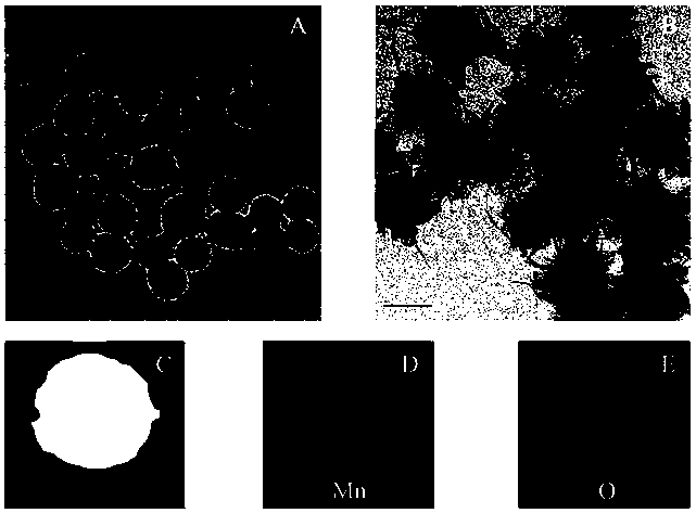
[0036] refer to figure 1 , 1mg / L of PDA@MnO 2 The sample aqueous solution was dropped on the copper grid, and the sample was completely dried for transmission electron microscope characterization. In the figure, A is a transmission electron microscope photo of PDA nanoparticles, and it can be seen that the particle size is about 100nm; B is PDA@MnO 2 The transmission electron microscope photo shows that 2-5nm MnO is reduced on the surface of the material. 2 The layer is monodisperse; Figures C, D, and E are the element mapping white field, manganese element, and oxygen element photos, showing that the surface of the material contains manganese element and oxygen element.
PUM
| Property | Measurement | Unit |
|---|---|---|
| Particle size | aaaaa | aaaaa |
| Particle size | aaaaa | aaaaa |
Abstract
Description
Claims
Application Information
 - R&D
- Intellectual Property
- Life Sciences
- Materials
- Tech Scout
- Unparalleled Data Quality
- Higher Quality Content
- 60% Fewer Hallucinations
Browse by: Latest US Patents, China's latest patents, Technical Efficacy Thesaurus, Application Domain, Technology Topic, Popular Technical Reports.
© 2025 PatSnap. All rights reserved.Legal|Privacy policy|Modern Slavery Act Transparency Statement|Sitemap|About US| Contact US: help@patsnap.com


