
Microfluidic chip imaging system for monitoring cell pharmacokinetics

A detection system and microfluidic technology, applied in tissue cell/virus culture devices, enzymology/microbiology devices, specific-purpose bioreactors/fermenters, etc. advanced questions

Inactive Publication Date: 2018-03-09
HENAN TIANSHENG TAIFENG PHARM TECH CO LTD
View PDF0 Cites 2 Cited by
  • Summary
  • Abstract
  • Description
  • Claims
  • Application Information

AI Technical Summary

Problems solved by technology

[0002] At present, the most commonly used molecular imaging technology is nuclear medicine imaging technology, especially the molecular imaging research of positron emission imaging PET is the most influential. The current positron emission imaging technology is limited to human body or small animal imaging and has a large equipment volume. , high cost, and difficult manipulation, etc., there is no application for in vitro imaging, that is, cell imaging of resected tissue slices

Method used


Image

  • Microfluidic chip imaging system for monitoring cell pharmacokinetics
  • Microfluidic chip imaging system for monitoring cell pharmacokinetics
  • Microfluidic chip imaging system for monitoring cell pharmacokinetics

Examples


Embodiment Construction

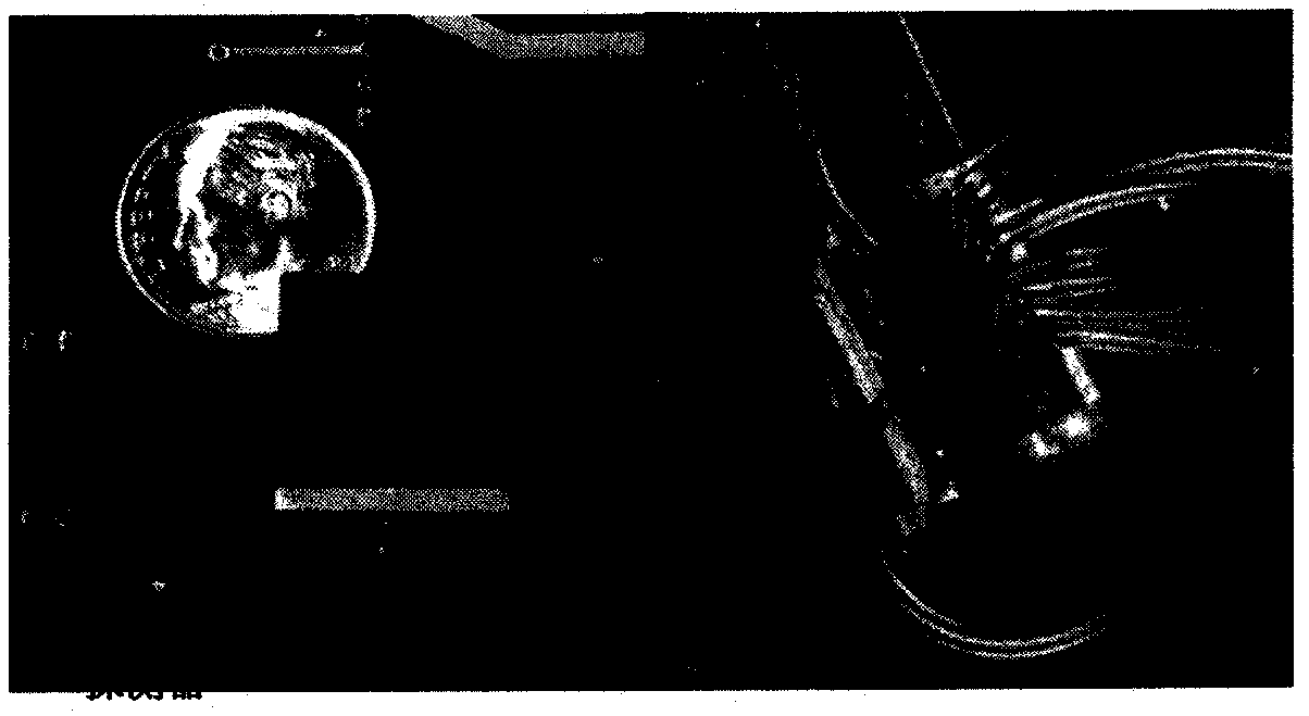
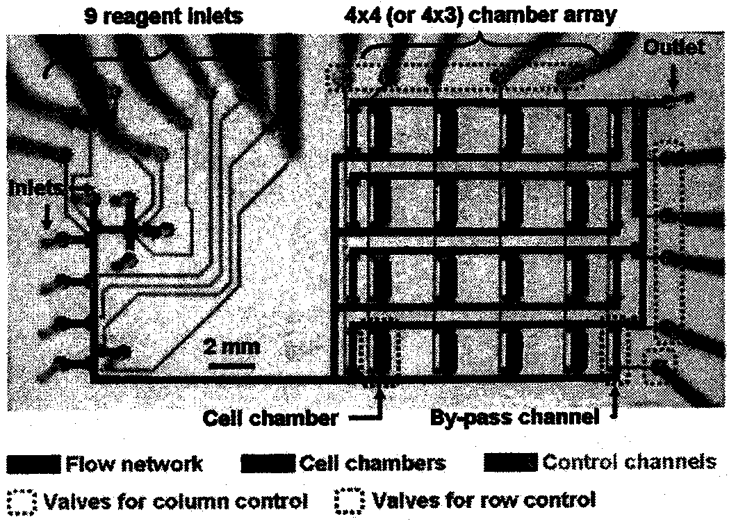
[0004] Such as figure 1 .The right figure shows the measurement of cell pairs Fluorodeoxyglucose [ 18 F] The absorption process of FDG: (1) cells are loaded into the culture grid (2) the [ 18 F] FDG solution is introduced (3) cell uptake[ 18 F]FDG (4) converts extracellular [ 18 F] The FDG solution is washed, and the detector only measures the intracellular [ 18 F] the concentration of FDG and reconstruct the image of the cell by detecting the positrons emitted from the cell.

[0005] illustrate:[ 18 F]FDG means Fluorodeoxyglucose . Glucose is one of the three major energy substances in the human body, and it will be detected by PET and form an image of the positron nuclide 18 F is labeled on glucose, which forms the [ 18 F] FDG; because [ 18 F] FDG can accurately reflect the glucose metabolism level of organs / tissues in the body, so it is currently the main imaging agent for PET-CT imaging; due to the vigorous metabolism of malignant tumor cells, the demand for gl...


PUM


Abstract

The invention relates to a microfluidic imaging system, specifically a high-precision sensitive detector and a microfluidic chip layer, which are applied in the fields of drug screening and precision medicine. It integrates functional modules such as multifunctional microvalve, positron detection, and image reconstruction analysis on the basis of existing microfluidic chips, and uses computers for unmanned automatic control, and develops a set that can be used in the development and research of modern biomedicine. It is a digital valve-controlled biomedical development microfluidic platform that integrates precise sample injection, chemical reaction synthesis, biological drug screening, cell culture and online drug effect analysis. Realize low-cost, high-throughput biopharmaceutical development, synthesis and screening, virus resistance screening, individualized drug tracking and other functions.

Description

technical field [0001] The invention relates to a microfluidic imaging system, specifically a high-precision sensitive detector and a microfluidic chip layer, which are applied in the fields of drug screening and precision medicine. Background technique [0002] At present, the most commonly used molecular imaging technology is nuclear medicine imaging technology, especially the molecular imaging research of positron emission imaging PET is the most influential. The current positron emission imaging technology is limited to human body or small animal imaging and has a large equipment volume. However, due to the disadvantages of high cost and difficult manipulation, there is no application for in vitro imaging, that is, cell imaging of resected tissue slices. The invention combines the microfluidic chip and the principle of positron emission imaging to construct an in vitro imaging system integrating cell culture, detection and imaging. Contents of the invention [0003] T...

Claims


Application Information

Patent Timeline
Patent Type & Authority Applications(China)
IPC IPC(8): C12M3/00C12M1/34
CPCG01N33/502
Inventor 沙薇
Owner HENAN TIANSHENG TAIFENG PHARM TECH CO LTD
Learn More