Microwave thermoacoustic early hepatoma detection device and method

A technology of early stage liver cancer and detection device, applied in the direction of organ motion/change detection, ultrasonic/sonic/infrasonic diagnosis, ultrasonic/sonic/infrasonic Permian technology, etc. Long continuous operation time, light weight and minimal thermal damage

Inactive Publication Date: 2018-03-13
SOUTH CHINA NORMAL UNIVERSITY
View PDF6 Cites 12 Cited by
  • Summary
  • Abstract
  • Description
  • Claims
  • Application Information

AI Technical Summary

Problems solved by technology

At present, the detection stage of liver cancer is mostly in the late stage, and the onset of liver cancer is rapid, which is not conducive to the treatment of patients, and has brought great pain to patients and their families.

Method used


Image

  • Microwave thermoacoustic early hepatoma detection device and method
  • Microwave thermoacoustic early hepatoma detection device and method
  • Microwave thermoacoustic early hepatoma detection device and method

Examples


Embodiment

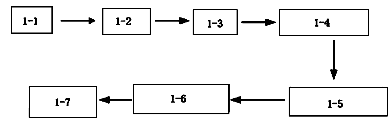
[0029] figure 1 It is a schematic diagram of the microwave thermoacoustic early liver cancer imaging detection device of the present invention, including a microwave trigger 1-1, a microwave generator 1-2, a transmitting antenna 1-3, an ultrasonic transducer 1-5, and a data acquisition card 1 connected in sequence -6 and computer 1-7, the microwave generator is triggered by the microwave trigger, and transmits pulsed microwaves to the liver of the human body to be measured through the transmitting antenna, and uses the thermoacoustic effect to excite and generate ultrasonic signals; the ultrasonic transducer converts the ultrasonic signals After being converted into an electrical signal, it is transmitted to the data acquisition card, and finally input to the computer, and the image is reconstructed by using the filter back projection algorithm and ultrasonic processing technology. In this embodiment, the pulse sequence of the microwave trigger is triggered by a computer. The...


PUM


Abstract

The invention relates to a microwave thermoacoustic early hepatoma detection device and method. The device comprises a microwave trigger, a microwave generator, a transmitting antenna, an ultrasonic transducer, a data acquisition card and a computer connected successively. The microwave generator transmits pulsed microwaves to the liver of the detected human body through the transmitting antenna under the triggering of the microwave trigger, and ultrasonic signals are generated by utilizing thermoacoustic effects; and the ultrasonic transducer converts the ultrasonic signals into electric signals, transmits the electric signals to the data acquisition card and finally inputs into the computer for image reconstruction by using a filter back projection algorithm and an ultrasonic processingtechnology. The detection device adopts a non-destructive detection technology and has the advantages of high contrast, high resolution, large visual field, easy adjustment and the like.

Description

technical field [0001] The invention belongs to the technical field of microwave thermoacoustic imaging, and in particular relates to a microwave thermoacoustic early liver cancer detection device and method. Background technique [0002] At present, the mortality rate of liver cancer in my country ranks third, second only to gastric cancer and esophageal cancer; the liver is the largest digestive organ in the human body, and damage to the liver will undoubtedly cause great harm to health. At present, the detection stage of liver cancer is mostly in the late stage, and the onset of liver cancer is rapid, which is not conducive to the treatment of patients, and has brought great pain to patients and their families. Early detection of liver cancer is of great help to the surgical resection and guidance of treatment of liver cancer, and can greatly improve the cure rate of liver cancer. [0003] The main tissue components of the liver are: water, protein, fat, carbohydrates, e...

Claims


Application Information

Patent Timeline
Patent Type & Authority Applications(China)
IPC IPC(8): A61B5/05A61B5/00A61B8/08
CPCA61B5/0033A61B5/0093A61B5/0507A61B5/4244A61B8/085A61B8/4444A61B8/4488A61B8/5215A61B8/5246A61B8/5261A61B2576/02
Inventor 计钟李亚平邢达杨思华
Owner SOUTH CHINA NORMAL UNIVERSITY
Why Eureka
Try Eureka