
Simulated thoracoscope simulation training device for surgery

A simulation training and thoracoscopic technology, applied in the field of medical equipment, can solve the problems of unavailable surgical training, operation training, unpredictable damage, and not being realistic enough, so as to improve basic skills and complex skills, shorten the learning curve of surgery, and facilitate training Effect

Pending Publication Date: 2018-03-13
THE FIRST AFFILIATED HOSPITAL OF MEDICAL COLLEGE OF XIAN JIAOTONG UNIV
View PDF7 Cites 12 Cited by
  • Summary
  • Abstract
  • Description
  • Claims
  • Application Information

AI Technical Summary

Problems solved by technology

Therefore, the operating direction is in a mirror image and lever relationship with the actual direction inside the video endoscope, and the operator's perception of space has obvious changes, which lead to a prolonged learning curve for endoscopic surgeons.
[0006] Second, one of the difficulties in thoracoscopic surgery is the continuous activity of the lungs and heart
[0007] Third, the main feature of thoracoscopic and endoscopic surgery is minimally invasive, but the corresponding risk is unpredictable injury during the operation (accidental accident)
The thoracoscopic simulator can only simulate basic operations such as cutting, separation, ligation, etc., but cannot simulate the physiological environment of the human body and the real surgical scene; the virtual thoracic mirror simulation system can simulate the surgical scene, but it is not realistic enough, and it can also simulate relatively complicated ones. Surgical environment, but lack of real-time force feedback system, real surgical training operation training, lack of the most important operation perception feedback during surgery, and high price, which limits its wide-scale promotion and use

Method used


Image

  • Simulated thoracoscope simulation training device for surgery
  • Simulated thoracoscope simulation training device for surgery
  • Simulated thoracoscope simulation training device for surgery

Examples


Embodiment

[0051] Such as image 3 as shown,

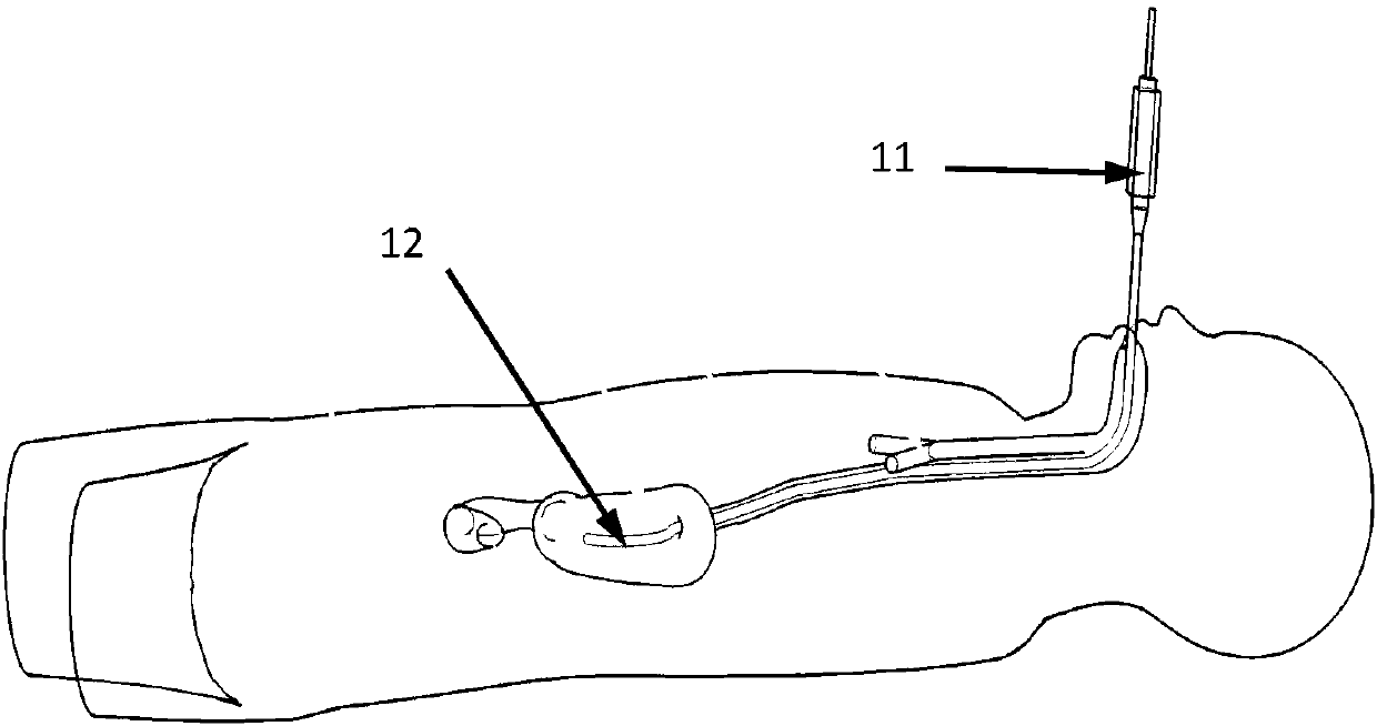
[0052] During the training process of simulated gastroscopic surgery, the trainee inserts the gastroscope directly from the simulated gastric tube set set on the simulator, holds the manipulation part with his left hand, and holds the mirror at a distance of 20 cm from the end of the mirror with his right hand, aiming the mirror at the oral cavity of the simulated person. Insert the end of the scope from the simulated esophageal tube set into the simulated stomach. During the whole process, the blood in the organs circulates by itself under the action of the circulation pump, and the mechanical ventilation device is used to simulate the human respiratory system. In this way, the intern can deeply experience the real operation process.

[0053] Such as Figure 4 as shown,

[0054] The operator uses the operating handle 15 of the bronchoscope to send the bronchoscope 14 sequentially through the simulated tracheal tube group, the simulated ...


PUM


Abstract

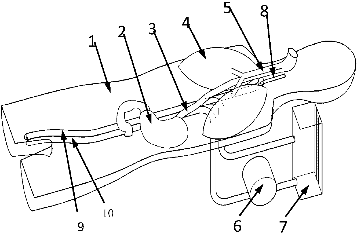
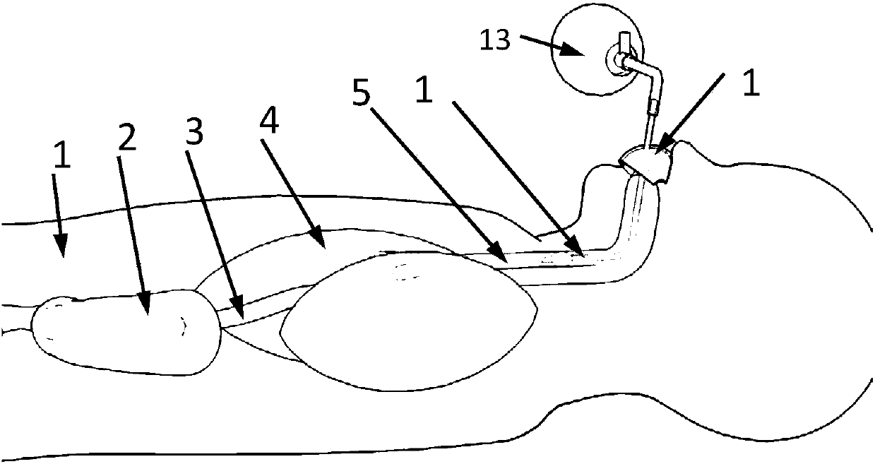
The invention relates to a simulated thoracoscope simulation training device for surgery. The device comprises a dummy man unit, a blood collection pool for collecting blood and connected with the dummy man unit, and a pump unit providing the perfusate circulation driving force. The dummy man unit comprises a dummy head and a dummy thoracoabdominal cavity unit connected with the dummy head. The dummy thoracoabdominal cavity unit consists of a front wall skin and a back skin that are separable and that are connected by a sealing buckle to form a hollow cavity provided therein with a dummy thoracic cavity and a dummy abdominal unit. The dummy thoracoabdominal cavity unit is also provided therein with an animal visceral organ group for simulation training. The animal visceral organ group includes a dummy lung and a dummy stomach. The dummy lung is disposed inside the dummy thoracic cavity. The dummy stomach is disposed inside the dummy abdominal cavity. The simulated thoracoscope surgerysimulation training device improves simulation training level of surgery and has low cost.

Description

technical field [0001] The invention belongs to the technical field of medical devices, and relates to a surgical training device for thoracoscope and multi-chamber endoscope (bronchoscope, mediastinoscope, gastroscope and enteroscope, etc.) with self-breathing function, in particular to a surgical simulation Thoracoscopic simulation training device. Background technique [0002] Endoscopic therapy is a technology that emerged after people solved the problem of thermal damage to tissues caused by light sources. At the end of the 19th century, more practical endoscopic instruments could be produced. In 1901, Kelling et al. reported laparoscopy using a cystoscope to examine the peritoneal cavity of dogs. In 1910, Jacobaeus, a brave Swedish professor of internal medicine, successfully applied this operation method to the human body for the first time, but due to the limitations of equipment and technology, thoracoscopic technology was only used for the diagnosis of pleural di...

Claims


Application Information

Patent Timeline
IPC IPC(8): G09B23/28G09B23/30
CPCG09B23/285G09B23/288G09B23/303
Inventor 张晓刚吕毅刘雯雁郑幸龙杨桓
Owner THE FIRST AFFILIATED HOSPITAL OF MEDICAL COLLEGE OF XIAN JIAOTONG UNIV
Learn More