Method for tissue evaluation of collagen at incisal edge of rectal cancer resection specimen

A collagen tissue and evaluation method technology, applied in the field of pathological identification, can solve the problems of affecting the healing of the anastomotic stoma, reducing the quality of life of the patient, and increasing the stenosis of the anastomotic stoma, achieving good scientific research and promotion value, avoiding serious complications, and improving the quality of life. Effect

Active Publication Date: 2018-04-20
NANFANG HOSPITAL OF SOUTHERN MEDICAL UNIV
View PDF3 Cites 0 Cited by
  • Summary
  • Abstract
  • Description
  • Claims
  • Application Information

AI Technical Summary

Problems solved by technology

[0003] However, while neoadjuvant chemotherapy and radiotherapy can reduce the local recurrence rate of rectal tumors, increase the rate of anus preservation, and prolong the survival of patients, the local tissue collagen hyperplasia caused by radiotherapy and chemotherapy will affect the healing of the anastomotic stoma, thereby increasing the incidence of postoperative complications. Possibility of complications such as anastomotic stenosis and anastomotic leakage
Among them, anastomotic leakage manifests as persistent high fever, paralytic ileus, acute peritonitis and other symptoms, and severe cases can be life-threatening. It is a common serious complication in rectal cancer and one of the main causes of death; anastomotic stenosis often causes The patient has poor defecation, frequent defecation, and even acute closed-loop intestinal obstruction, which reduces the patient's quality of life

Method used


Image

  • Method for tissue evaluation of collagen at incisal edge of rectal cancer resection specimen
  • Method for tissue evaluation of collagen at incisal edge of rectal cancer resection specimen
  • Method for tissue evaluation of collagen at incisal edge of rectal cancer resection specimen

Examples


Embodiment 1

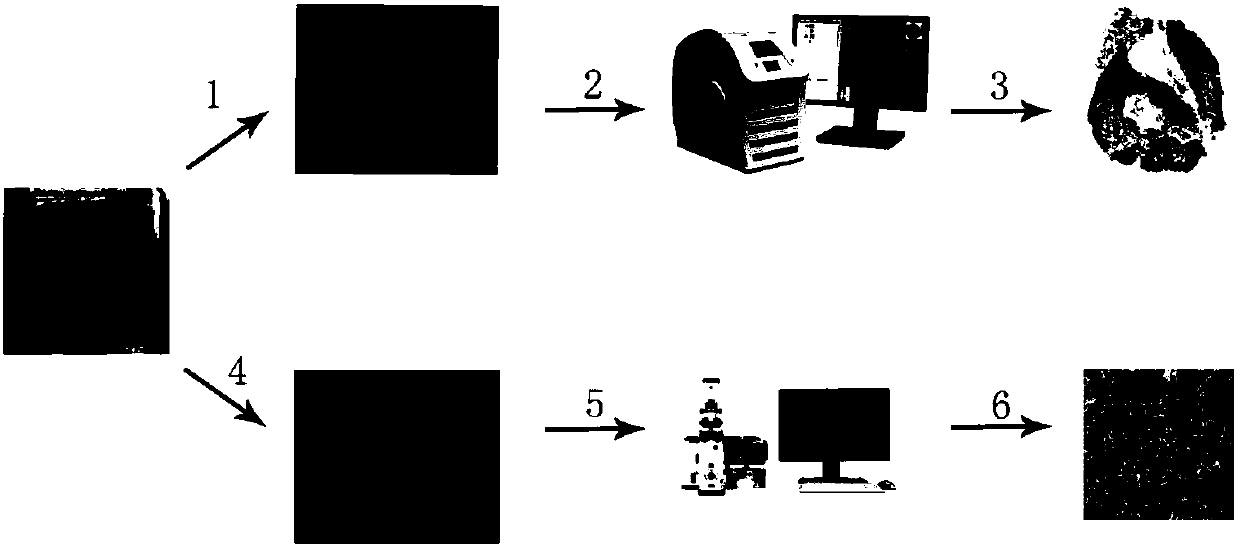
[0060] Such as figure 1 As shown, a method for evaluating collagen tissue at the margin of a rectal cancer resection specimen, the method specifically includes the following steps:

[0061] (a) Sample preparation: the resection margins of the rectal cancer specimens were taken out and then routinely prepared with wax blocks. Two tissue slices were continuously cut from each piece of tissue. The thickness of the tissue slices was 3 μm, and one was taken for pathological Masson staining. The Masson-stained sections were obtained, and the remaining tissue sections were infused into the submucosa of the tissue and placed in a -86°C refrigerator for refrigerated storage as the multiphoton imaging sections to be tested;

[0062] (b) Masson slice scanning: the Masson-stained slices were imaged as a whole by a slice scanner, the scanning imaging magnification was 15 times, and the resolution was 0.50 μm / image. After the imaging was completed, the tissue submucosa was marked by softwar...

Embodiment 2

[0066] A method for evaluating collagen tissue at the margin of a rectal cancer resection specimen, the method specifically comprising the following steps:

[0067] (a) Sample preparation: the resection margins of the rectal cancer specimens were taken out and then routinely prepared with wax blocks. Three tissue sections were continuously cut from each piece of tissue. The thickness of the tissue sections was 8 μm, and one piece was taken for pathological Masson staining. The Masson-stained sections were obtained, and the remaining tissue sections were infused into the submucosa of the tissue and placed in a -86°C refrigerator for refrigerated storage as the multiphoton imaging sections to be tested;

[0068] (b) Masson slice scanning: the Masson-stained slices were imaged as a whole through a slice scanner, the scanning imaging magnification was 25 times, and the resolution was 0.50 μm / image. After the imaging was completed, the tissue submucosa was marked by software;

[00...

Embodiment 3

[0072] A method for evaluating collagen tissue at the margin of a rectal cancer resection specimen, the method specifically comprising the following steps:

[0073] (a) Sample preparation: the resection margins of the rectal cancer specimens were taken out and then routinely prepared with wax blocks. Two tissue sections were continuously cut from each piece of tissue. The thickness of the tissue sections was 5 μm, and one was taken for pathological Masson staining. The Masson stained section was prepared, and the other tissue section was infused with the tissue submucosa and placed in a -86°C refrigerator for refrigerated storage as the multiphoton imaging section to be tested;

[0074] (b) Masson slice scanning: the Masson-stained slices were imaged as a whole through a slice scanner, the scanning imaging magnification was 20 times, and the resolution was 0.50 μm / image. After the imaging was completed, the tissue submucosa was marked by software;

[0075] (c) Marking the imag...


PUM

PropertyMeasurementUnit
thicknessaaaaaaaaaa

Abstract

The invention provides a method for tissue evaluation of collagen at the incisal edge of a rectal cancer resection specimen, belonging to the technical field of pathological differentiation. Accordingto the method, tissue evaluation is carried out on collagen at the incisal edge of a rectal cancer resection specimen obtained after neoadjuvant therapy of the rectal cancer in virtue of a combination of Masson dyeing and multiphoton imaging analysis, so clinical workers are allowed to learn about the local fibration degree of the rectum tissue of patients and the healing status of anastomotic parts; and thus, reference is provided for clinical doctors in selection of surgical methods and in subsequent diagnosis and treatment, better individual-based treatment schemes are provided for patients, and the purposes of improving the living quality of patients and maximally avoiding severe complications are eventually achieved. The method provided by the invention is reasonable in design, mature in technology and easy to practice and has good clinical application prospects and high promotion value in scientific research.

Description

technical field [0001] The invention relates to the technical field of pathological identification, in particular to a method for evaluating collagen tissue at the cutting edge of a rectal cancer resection specimen. Background technique [0002] Rectal cancer is one of the common gastrointestinal tumors. According to the World Health Organization International Cancer Research Center (IARC), there were about 1.36 million new cases of rectal cancer in the world in 2012, ranking second among malignant tumors after lung cancer, liver cancer and gastric cancer. No. 4. In recent years, with the deepening of modern treatment concepts for rectal tumors, the efficacy of neoadjuvant chemoradiotherapy for locally advanced rectal cancer has been confirmed. Neo-adjuvant chemoradiotherapy (Neo-CRT) refers to preoperative radiotherapy Chemotherapy measures. Because the blood supply of the rectal tumor is sound before operation, the hypoxic tumor cells are few, and it is sensitive to radi...

Claims


Application Information

Patent Timeline
Patent Type & Authority Applications(China)
IPC IPC(8): G01N21/63G01N1/28
CPCG01N1/28G01N21/63
Inventor 严俊蒋伟许春辉李国新徐硕瑀刘张苑珠陈德鑫李凯严博韬
Owner NANFANG HOSPITAL OF SOUTHERN MEDICAL UNIV
Why Eureka
Try Eureka