Evaluation method of collagen tissue at the resection margin of rectal cancer resection specimens
An evaluation method and collagen tissue technology, applied in the field of pathological identification, can solve problems such as affecting the healing of the anastomotic stoma, reducing the quality of life of the patient, and increasing the stenosis of the anastomotic stoma, achieving good scientific research and promotion value, avoiding serious complications, and improving the quality of life. Effect
- Summary
- Abstract
- Description
- Claims
- Application Information
AI Technical Summary
Problems solved by technology
Method used
Image
Examples
Embodiment 1
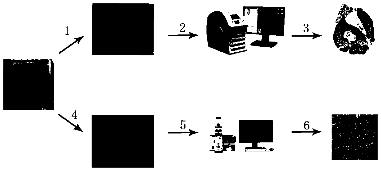
[0060] Such as figure 1 As shown, a method for evaluating collagen tissue at the margin of a rectal cancer resection specimen, the method specifically includes the following steps:
[0061] (a) Sample preparation: the resection margins of the rectal cancer specimens were taken out and then routinely prepared with wax blocks. Two tissue slices were continuously cut from each piece of tissue. The thickness of the tissue slices was 3 μm, and one was taken for pathological Masson staining. The Masson-stained sections were obtained, and the remaining tissue sections were infused into the submucosa of the tissue and placed in a -86°C refrigerator for refrigerated storage as the multiphoton imaging sections to be tested;
[0062] (b) Masson slice scanning: the Masson-stained slices were imaged as a whole by a slice scanner, the scanning imaging magnification was 15 times, and the resolution was 0.50 μm / image. After the imaging was completed, the tissue submucosa was marked by softwar...
Embodiment 2
[0066] A method for evaluating collagen tissue at the margin of a rectal cancer resection specimen, the method specifically comprising the following steps:
[0067] (a) Sample preparation: the resection margins of the rectal cancer specimens were taken out and then routinely prepared with wax blocks. Three tissue sections were continuously cut from each piece of tissue. The thickness of the tissue sections was 8 μm, and one piece was taken for pathological Masson staining. The Masson-stained sections were obtained, and the remaining tissue sections were infused into the submucosa of the tissue and placed in a -86°C refrigerator for refrigerated storage as the multiphoton imaging sections to be tested;
[0068] (b) Masson slice scanning: the Masson-stained slices were imaged as a whole through a slice scanner, the scanning imaging magnification was 25 times, and the resolution was 0.50 μm / image. After the imaging was completed, the tissue submucosa was marked by software;
[00...
Embodiment 3
[0072] A method for evaluating collagen tissue at the margin of a rectal cancer resection specimen, the method specifically comprising the following steps:
[0073] (a) Sample preparation: the resection margins of the rectal cancer specimens were taken out and then routinely prepared with wax blocks. Two tissue sections were continuously cut from each piece of tissue. The thickness of the tissue sections was 5 μm, and one was taken for pathological Masson staining. The Masson stained section was prepared, and the other tissue section was infused with the tissue submucosa and placed in a -86°C refrigerator for refrigerated storage as the multiphoton imaging section to be tested;
[0074] (b) Masson slice scanning: the Masson-stained slices were imaged as a whole through a slice scanner, the scanning imaging magnification was 20 times, and the resolution was 0.50 μm / image. After the imaging was completed, the tissue submucosa was marked by software;
[0075] (c) Marking the imag...
PUM
| Property | Measurement | Unit |
|---|---|---|
| thickness | aaaaa | aaaaa |
Abstract
Description
Claims
Application Information
 - R&D
- Intellectual Property
- Life Sciences
- Materials
- Tech Scout
- Unparalleled Data Quality
- Higher Quality Content
- 60% Fewer Hallucinations
Browse by: Latest US Patents, China's latest patents, Technical Efficacy Thesaurus, Application Domain, Technology Topic, Popular Technical Reports.
© 2025 PatSnap. All rights reserved.Legal|Privacy policy|Modern Slavery Act Transparency Statement|Sitemap|About US| Contact US: help@patsnap.com


