Single-scanning magnetic resonance quantitative T2 imaging reconstruction method based on residual network

A magnetic resonance imaging and single-scanning technology, which is applied in the fields of nuclear magnetic resonance analysis, 2D image generation, image generation, etc., can solve the problem of difficult separation of four overlapping echo signals, so as to improve accuracy and expand the measurement range , the effect of large measurement range

Active Publication Date: 2018-05-08
XIAMEN UNIV
View PDF5 Cites 19 Cited by
  • Summary
  • Abstract
  • Description
  • Claims
  • Application Information

AI Technical Summary

Problems solved by technology

However, it is difficult to separate the four overlapping echo signals by conventional methods

Method used


Image

  • Single-scanning magnetic resonance quantitative T2 imaging reconstruction method based on residual network
  • Single-scanning magnetic resonance quantitative T2 imaging reconstruction method based on residual network
  • Single-scanning magnetic resonance quantitative T2 imaging reconstruction method based on residual network

Examples


specific Embodiment

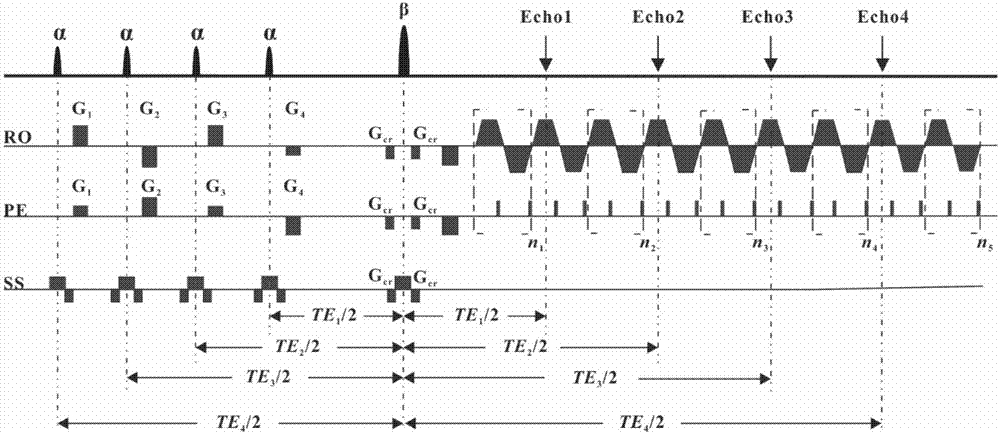
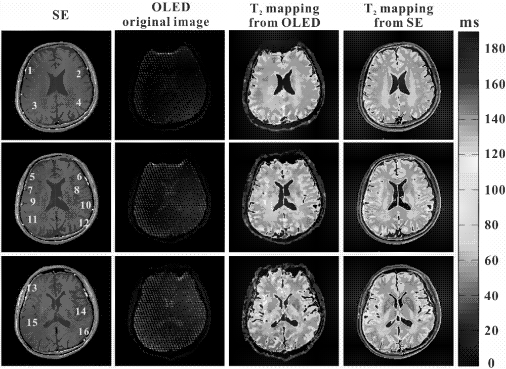
[0081] Quantification of T using residual network-based single-scan MR 2 The imaging reconstruction method carried out human brain experiments to verify the feasibility of the present invention. The experiment was carried out under the human body nuclear magnetic resonance 3T imager. On the operating table of the magnetic resonance imager, open the corresponding operating software in the imager, first locate the region of interest of the imaging object, and then perform tuning, shimming, power and frequency correction. In order to evaluate the validity of the image obtained by this method, SE imaging experiments were carried out in the same environment as a comparison. Then import the compiled OLED imaging sequence (such as figure 1 As shown), according to the specific experimental situation, set each parameter of the pulse sequence. The experimental parameters of this embodiment are set as follows: the imaging field of view FOV is 22cm × 22cm, the excitation time of the 15°...


PUM


Abstract

The invention discloses a single-scanning magnetic resonance quantitative T2 imaging reconstruction method based on a residual network and relates to magnetic resonance imaging methods. According to the method, four small-angle excitation pulses with the same deflection angle are utilized, and a period of evolution time is available after each excitation pulse, so that transverse relaxation time T2 of each echo signal is different; a shift gradient of a frequency coding dimension and a phase coding dimension is added after each excitation pulse, so that the positions of the signals generated by different excitation pulses in a k space are different; multiple echo signals with different transverse relaxation time are obtained in one time of sampling; then the sampling signals are input intothe trained residual network after being subjected to normalization, zero-setting and fast Fourier transformation for reconstruction to obtain a quantitative T2 image; training data of the residual network comes from simulation data; and a template is randomly generated first, then an input image of the network is obtained by simulating experiment environment sampling, the template is used as a tag, and a mapping relation between the input image and an output image is obtained through training.

Description

technical field [0001] The invention relates to a magnetic resonance imaging method, in particular to a single-scan magnetic resonance quantitative T method based on a residual network 2 Imaging (T 2 mapping) reconstruction method. Background technique [0002] Magnetic resonance parametric imaging (eg, T 1 Imaging, T 2 Imaging and T 2 * imaging) can be readily used to provide quantitative information for characterizing specific tissue properties [1] . Quantitative imaging has many outstanding features, the most obvious of which is the ability to eliminate effects independent of tissue properties, such as operator dependence, differences in scanning parameters, spatial variations in magnetic field, and image scaling. [2] . Quantitative T 2 Imaging has a wide range of clinical applications involving early diagnosis of neurodegenerative diseases [3] , liver iron overload measurement [4] , myocardial infarction assessment [5] , Quantification of labeled cells [6] ,...

Claims


Application Information

Patent Timeline
Patent Type & Authority Applications(China)
IPC IPC(8): G06T11/00G01N24/08
CPCG01N24/08G06T11/003G06T2207/10088G06T2207/20056G06T2207/20081G06T2211/416
Inventor 蔡淑惠张俊蔡聪波廖璞曾坤
Owner XIAMEN UNIV
Why Eureka
Try Eureka