Preparation method of composite cavity microfibers based on micro-fluidic technology

A microfluidic technology and microfiber technology, which is applied in the field of cavity composite microfiber preparation, can solve the problems of unstable cell adhesion, limited application, unfavorable sodium alginate, etc., and achieve attachment and growth, enhanced stability, Effect of promoting adhesion and growth

Active Publication Date: 2018-06-12
DALIAN INST OF CHEM PHYSICS CHINESE ACAD OF SCI
View PDF2 Cites 13 Cited by
  • Summary
  • Abstract
  • Description
  • Claims
  • Application Information

AI Technical Summary

Problems solved by technology

However, sodium alginate is not conducive to cell adhesion and is

Method used


Image

  • Preparation method of composite cavity microfibers based on micro-fluidic technology
  • Preparation method of composite cavity microfibers based on micro-fluidic technology
  • Preparation method of composite cavity microfibers based on micro-fluidic technology

Examples


Embodiment 1

[0075] A method for preparing composite cavity microfibers based on microfluidic technology. In the process of preparing cavity microfibers, a modified material that can promote cell adherent growth is introduced into the inner cavity of the microfibers, and the cavity is formed At the same time, the modified material is attached to the cavity to form a modified coating, which can promote the adhesion and culture of cells in the later stage.

[0076] The microfluidic chip is composed of upper and lower layers of chips, both of which are made of polydimethylsiloxane; the chip has at least three parallel channel inlets and one general outlet; the microfluidic technology-based compound The requirements for the preparation method of hollow microfibers are as follows:

[0077] Lay the microfluidic chip flat, submerge the outlet of the chip in the receiving liquid, connect the syringe to the inlet of the microfluidic chip, and use the syringe pump to push the syringe to form a multi...

Embodiment 2

[0110] The content of this embodiment is basically the same as that of Embodiment 1, and its difference mainly lies in:

[0111] The microfluidic chip has a three-channel structure; compared with the microfluidic chip with a four-channel structure, the microfluidic chip with a three-channel structure has three independent sample inlets and three independent channels, and the modified fluid and The inert fluids are mixed together and merged into a central fluid. The channels from inside to outside are the mixture of inert fluid and modified fluid (fluid 1), the sample fluid (fluid 2) and the sheath flow fluid (fluid 3).

[0112] And when the microfluidic chip is a three-channel structure, the sizes of the three channels from small to large are as follows: the channel size of the inert fluid and the cavity modification fluid mixture < the channel size of the sample fluid < the channel size of the sheath fluid; The channel size of the sample fluid is larger than the channel size ...

Embodiment 3

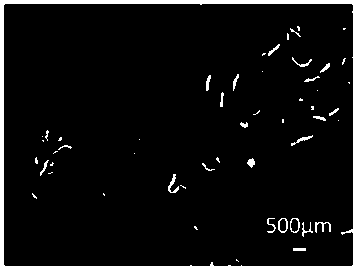
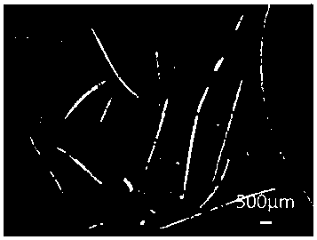
[0114] The chip used was a PDMS chip, which was sealed after oxygen plasma surface treatment, and then passed through perfluorinated liquid for 20 minutes to modify the surface of the channel. Vacuum dry the perfluorinated liquid and dry it in an oven at 80°C for later use. The chip structure diagram is as follows figure 1 shown.

[0115] 2% sodium alginate (viscosity 240mPa·s) containing 0.85% NaCl is used as sample fluid, 3% methylcellulose (viscosity 4000cP) is used as inert fluid, 5% chitosan ( Viscosity>400mPa s) as modified fluid, 1.1%CaCl containing 3% sucrose 2as a sheath fluid. The modification fluid can also be 5% chitosan and 3% methylcellulose mixed in different proportions to obtain methylcellulose solutions containing different chitosan concentrations (1%, 3%). Sterilize the methylcellulose at high temperature for half an hour, and then put it into a 4°C refrigerator to dissolve it for later use. Filter the sodium alginate solution with a 0.22 μm sterile fil...


PUM

PropertyMeasurementUnit
Viscosityaaaaaaaaaa
Viscosityaaaaaaaaaa
Viscosityaaaaaaaaaa

Abstract

The invention relates to a preparation method of composite cavity microfibers based on a micro-fluidic technology. The preparation method is characterized in that in the preparation process of cavitymicrofibers, a modified material capable of promoting the cell wall adhesion growth is introduced in a microfiber inner cavity; while the cavity is formed, the modified material is adhered onto the cavity to form a modified coating layer, so that a promoting effect on late cell adhesion and culture is provided. By utilizing the micro-fluidic technology, a micron-sized channel capable of generatinga coaxial laminar flow pattern is formed, the flow pattern control on a sample fluid is realized, and the sample fluid is finally solidified into a micron-sized hollow fiber material with a specificinner coating structure. The microfiber material can simulate a microstructure in a human body tissue, and a new method and a new concept are provided for tissue engineering and organ regeneration. The preparation method provided by the invention is simple and reliable to operate, high in efficiency and excellent in technical effect; convenient conditions are provided for the modification of the microfibers; an internal finish coating is uniform, stable, simple and controllable to facilitate the cell wall adhesion growth.

Description

technical field [0001] The invention relates to the field of preparation method and application technology of hollow composite microfibers based on microfluidic chip technology, and in particular provides a preparation method of composite hollow microfibers based on microfluidic technology. Background technique [0002] In the prior art, organ transplantation is an ideal method for treating organ failure, but problems such as organ supply shortage, immune rejection, and ethical disputes seriously restrict its clinical application. Therefore, tissue engineering, as a new way to construct implantable organs, has become an important development direction for the treatment of organ failure. Tissue engineering mainly uses biocompatible scaffold materials to combine with cells to prepare transplantable engineered tissues with cell functions. The purpose of organ shortage [1,2]. Therefore, in the construction process of tissue engineering, it is particularly important to simulate...

Claims


Application Information

Patent Timeline
IPC IPC(8): D01F8/18D01F8/02D01F11/00D01F11/02C12N5/071C12N5/09
CPCC12N5/0602C12N5/0693C12N2533/30C12N2533/32C12N2533/40C12N2533/52C12N2533/54C12N2533/72C12N2533/74C12N2533/76C12N2533/78C12N2533/80D01F8/02D01F8/18D01F11/00D01F11/02
Inventor 秦建华刘慧王亚清于跃
Owner DALIAN INST OF CHEM PHYSICS CHINESE ACAD OF SCI
Why Eureka
Try Eureka