Tumor volume intelligent sketching method and device
A volume and tumor technology, applied in the field of image analysis, can solve the problems of reducing the reliability of treatment, affecting the accuracy of tumor volume delineation, affecting the efficacy of radiotherapy, etc., to reduce the cost of manual delineation, ensure reliability, and improve accuracy.
- Summary
- Abstract
- Description
- Claims
- Application Information
AI Technical Summary
Problems solved by technology
Method used
Image
Examples
Embodiment 1
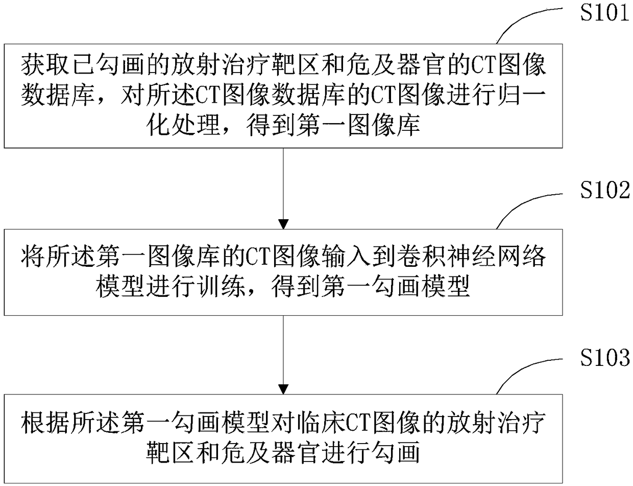
[0055] see figure 1 Provides a schematic diagram of the implementation process of an embodiment of the intelligent tumor volume delineation method, detailed as follows:
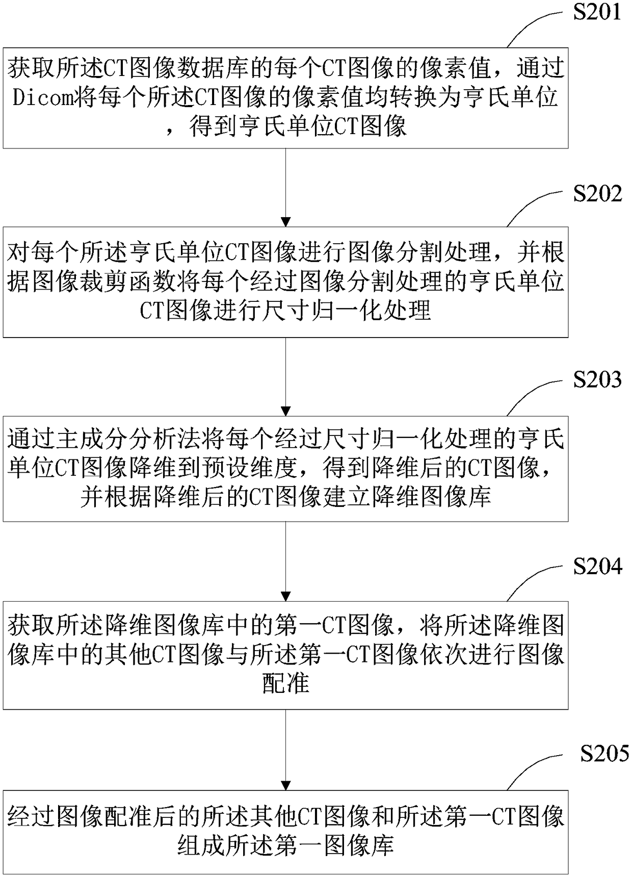
[0056] Step S101 , acquiring a CT image database of the delineated tumor volume, and performing normalization processing on the CT images in the CT image database to obtain a first image database.
[0057] Wherein, the CT image database of the delineated tumor volume in this embodiment is the CT image of the tumor volume accurately delineated by doctors obtained from a regular hospital. Preferably, the number of CT images in the CT image database is at least 300.
[0058] Optionally, before performing normalization processing on the CT images in the CT image database, the method may further include: performing image enhancement processing on the CT images in the CT image database. The image enhancement process is to highlight the tumor volume in the CT image, highlighting the feature information of the tumo...
Embodiment 2
[0126] Corresponding to the method for intelligently delineating the tumor volume described in the first embodiment above, Figure 8 shows a structural block diagram of the tumor volume intelligent delineation device in the second embodiment of the present invention. For ease of description, only the parts related to this embodiment are shown.
[0127] The device includes: a normalization module 110 , a model generation module 120 and a drawing module 130 .
[0128] The normalization module 110 is configured to obtain a CT image database of the delineated tumor volume, perform normalization processing on the CT images of the CT image database, and obtain a first image database.
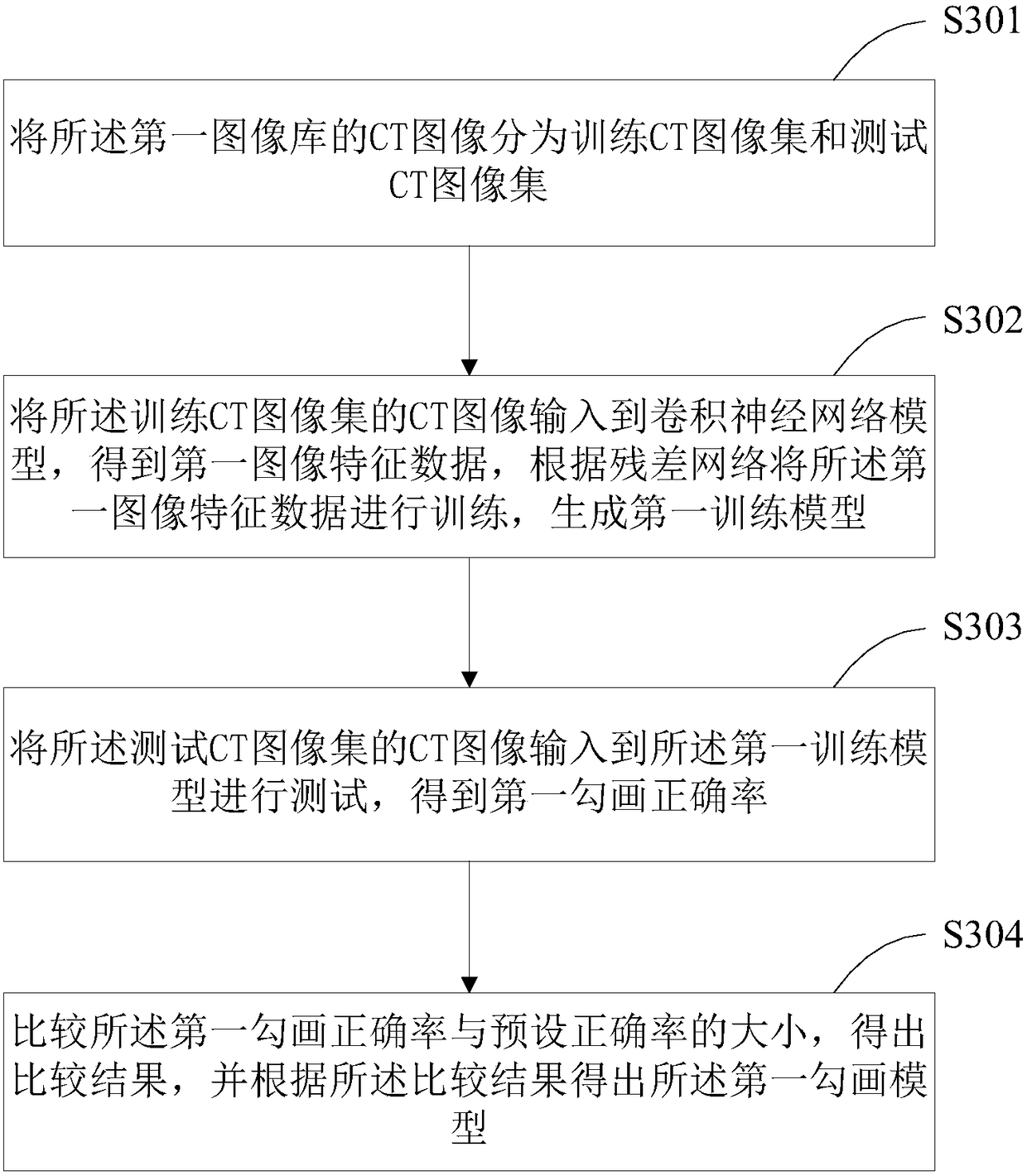
[0129] The model generating module 120 is configured to input the CT images of the first image library into the convolutional neural network model for training, and obtain a trained first delineation model.
[0130] The delineation module 130 is used for delineating the tumor volume of the clinical CT...
Embodiment 3
[0135] Figure 9 It is a schematic diagram of a terminal device 100 for intelligently delineating tumor volumes provided by Embodiment 3 of the present invention. Such as Figure 9 As shown, the terminal device 100 for intelligent tumor volume delineation in this embodiment includes: an image display device 150, a processor 160, a memory 170, and a computer stored in the memory 170 and capable of running on the processor 160 The program 171 is, for example, the program of the intelligent delineation method for tumor volume. When the processor 160 executes the computer program 171, it realizes the steps in the above embodiment of the method for intelligently delineating the tumor volume, for example figure 1 Steps 101 to 103 are shown. Alternatively, when the processor 160 executes the computer program 171, it realizes the functions of the modules / units in the above-mentioned device embodiments, for example Figure 8 The functions of modules 110 to 140 are shown.
[0136] ...
PUM
 Abstract
Description
Claims
Application Information
 - R&D
- Intellectual Property
- Life Sciences
- Materials
- Tech Scout
- Unparalleled Data Quality
- Higher Quality Content
- 60% Fewer Hallucinations
Browse by: Latest US Patents, China's latest patents, Technical Efficacy Thesaurus, Application Domain, Technology Topic, Popular Technical Reports.
© 2025 PatSnap. All rights reserved.Legal|Privacy policy|Modern Slavery Act Transparency Statement|Sitemap|About US| Contact US: help@patsnap.com


