Kit for quantitatively detecting myeloperoxidase, and preparation method thereof
A technology for quantitative detection of myeloperoxidase, which is applied in the field of kits and preparations for quantitative detection of myeloperoxidase, can solve the problems of poor specificity and low sensitivity, achieve high specificity, high sensitivity, and ensure sensitivity Effect
- Summary
- Abstract
- Description
- Claims
- Application Information
AI Technical Summary
Problems solved by technology
Method used
Image
Examples
Embodiment 1
[0052] Example 1 Preparation of MPO Acridine Lipid-labeled Monoclonal Antibody
[0053] 1) Weigh an appropriate amount of MPO monoclonal antibody and use 0.05mol / L pH 9.5 carbonate buffer (CB) to adjust to 1mg / mL;
[0054] 2) Acridine lipid and antibody are calculated at 1:15 molar ratio, dissolved in DMF, mixed, and reacted at room temperature for 30 minutes;
[0055] 3) Transfer the reaction solution to a dialysis bag (molecular weight cut-off 8000-12000), and dialyze with 0.01M PBS with pH 7.2 for 24 hours;
[0056] 4) Add an equal volume of glycerin and 0.1% PC-300.
[0057] The acridinium ester marker may be an acridone compound, an acridine sulfonamide compound or acridinyl aminoacetic acid.
Embodiment 2
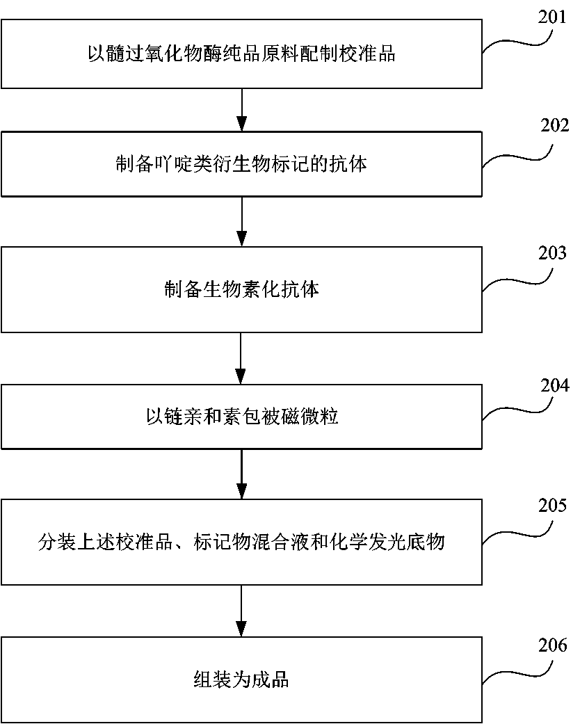
[0058] Example 2 Preparation of the MPO quantitative assay kit of the present invention
[0059] 1. Preparation of acridine lipid antibody
[0060] 1. Acridine-labeled monoclonal antibody diluent Buffer A:
[0061] Buffer A: 0.01-0.1M PBS pH 7.2, 0.1%-1%BSA, 0.2% PC-300
[0062] 2. Select the concentration of acridine-labeled monoclonal antibody:
[0063] The working concentration of acridinium-labeled monoclonal antibody is greater than 0.45 μg / mL.
[0064] 2. Preparation of MPO calibrator
[0065] Dilute the MPO antigen with 0.01M PBS pH 7.2, 0.5% BSA diluent, and prepare five kinds of calibrator with standard concentration of 0, 5, 25, 100, 500, 2000ng / mL.
[0066] 3. Preparation of antibodies labeled with acridine derivatives:
[0067] 1) Weigh an appropriate amount of MPO monoclonal antibody, and use 0.05mol / L pH 9.5 CB to adjust to 1mg / mL;
[0068] 2) Acridine derivatives and antibodies are calculated at 1:15 molar ratio, dissolved in DMF, mixed, and reacted at roo...
Embodiment 3
[0094] Example 3 Buffer Buffer
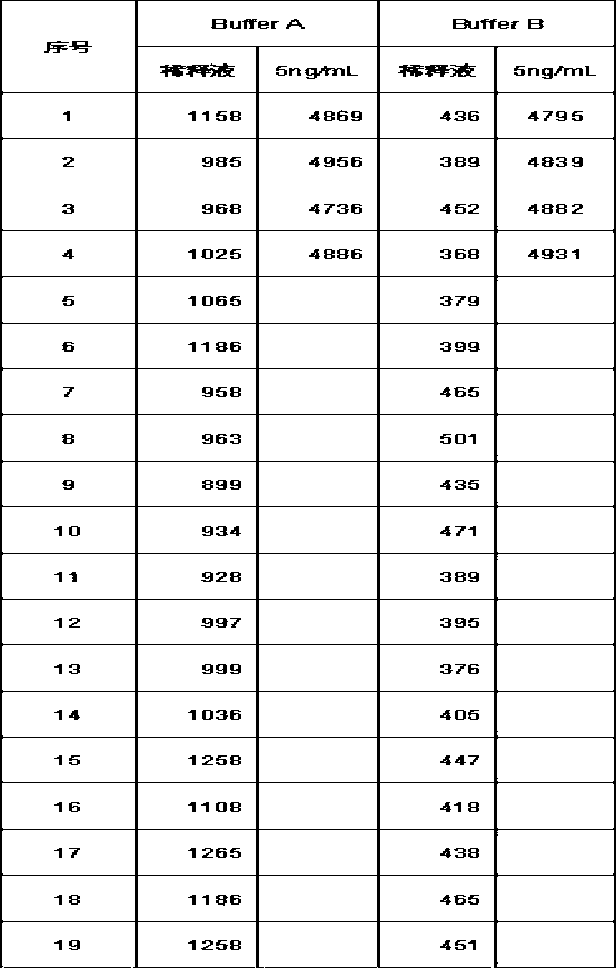
[0095] As an important component of the present invention, the buffer is a buffer for diluting biotinylated antibodies, antibodies labeled with acridine derivatives, and streptavidin-coated magnetic particles, using a combined formula of BSA, trehalose and casein, Improve the non-specific physical adsorption of immunoglobulin by the magnetic particles, further reduce the specific binding, and improve the analytical sensitivity of the reagent.
[0096] Buffer A: 0.01-0.1M PBS pH 7.2, mass ratio 0.1%-1%BSA, volume ratio 0.2% PC-300;
[0097] Buffer B: 0.01-0.1M PBS pH 7.2, mass ratio 0.1%-1%BSA, mass ratio 0.1%-5% trehalose, mass ratio 0.5%-1% casein, volume ratio 0.2% PC-300.
[0098] Analytical Sensitivity=2*(0 value RLU SD)*5 / (Calibrator RLU mean - 0 value RLU mean)
[0099]
[0100] Use Buffer B as the diluent of acridinium ester-MPO antibody conjugate (AE-MPO) 2#, AE-MPO4# (2#, 4# are antibody numbers), magnetic particle-streptavidin (M...
PUM
| Property | Measurement | Unit |
|---|---|---|
| Analytical sensitivity | aaaaa | aaaaa |
| Sensitivity | aaaaa | aaaaa |
Abstract
Description
Claims
Application Information
 - R&D
- Intellectual Property
- Life Sciences
- Materials
- Tech Scout
- Unparalleled Data Quality
- Higher Quality Content
- 60% Fewer Hallucinations
Browse by: Latest US Patents, China's latest patents, Technical Efficacy Thesaurus, Application Domain, Technology Topic, Popular Technical Reports.
© 2025 PatSnap. All rights reserved.Legal|Privacy policy|Modern Slavery Act Transparency Statement|Sitemap|About US| Contact US: help@patsnap.com


