Cerebral cortex structure, cerebral blood vessel, intracerebral lesion 3D printing model and preparation method thereof

A 3D printing, cerebral cortex technology, applied in the medical field, can solve the problems of incomplete eradication, wrong surgical judgment, preoperative planning errors, etc., to achieve the effect of improving clinical diagnosis and treatment skills, reducing surgical risks, and increasing benefits

Inactive Publication Date: 2018-09-14
RENJI HOSPITAL AFFILIATED TO SHANGHAI JIAO TONG UNIV SCHOOL OF MEDICINE
View PDF1 Cites 9 Cited by
  • Summary
  • Abstract
  • Description
  • Claims
  • Application Information

AI Technical Summary

Problems solved by technology

[0003] Craniocerebral surgery is still performed clinically based on two-dimensional (2-Dimensional, 2D) imaging and simulated three-dimensional (3-Dimensional, 3D) images for three-dimensional space imagination, although there are many clinically available commercial navigation and open source software It can be simulated and displayed in 3D on the screen, but because the structure of the lesion is deep and closely related to the surrounding cerebral blood vessels, slight changes in body and head position during the operation cannot be accurately responded to on the screen, and the relationship between some blood vessels and brain sulci is false on the screen. The 3D display is very different from the real situation ( figure 1 ), the situation that leads to surgical judgment errors cannot be completely eradicated
[0004] For example, the inventor used Brain-LAB software to perform 3D reconstruction on 300 epilepsy patients through preoperative MRI and magnetic resonance angiography (magnetic resonance venography, MRV) ( figure 1 A) Intraoperative navigation is performed by fusing blood vessel information with the three-dimensional model of the cerebral cortex to obtain data. This technology greatly improves the accuracy of positioning and is conducive to clinical promotion and application. However, in actual operation, the inventors found that this There is still a certain error between this method and the preoperative plan. During the operation, it is necessary to judge the distance between the brain sulcus or gyrus and the blood vessel. Misjudgment will often lead to unsatisfactory surgical results, and serious complications will result if there is a slight carelessness.

Method used


Image

  • Cerebral cortex structure, cerebral blood vessel, intracerebral lesion 3D printing model and preparation method thereof
  • Cerebral cortex structure, cerebral blood vessel, intracerebral lesion 3D printing model and preparation method thereof
  • Cerebral cortex structure, cerebral blood vessel, intracerebral lesion 3D printing model and preparation method thereof

Examples


Embodiment 1

[0042] Step 1. Select patients who are clearly diagnosed as intractable epilepsy at the Renji Hospital Affiliated to Shanghai Jiao Tong University School of Medicine, and undergo strict evaluation for surgical resection (with the consent of the patient or guardian, and signed informed consent); after CT, MRI , DTI, and CTA examinations to obtain patient imaging data; the acquisition of cranial imaging data is very important, and low-resolution data will lead to errors between the generated model and the real brain anatomy.

[0043] Head CT 1mm axial thin-slice scan; magnetic resonance with no spacing axial, coronal and sagittal Flair sequence and T1 axial, coronal, sagittal sequence 1mm thin scan, parallel DTI and vascular CTA examination; all All imaging data were output in DICOM format through our hospital's PACS software system.

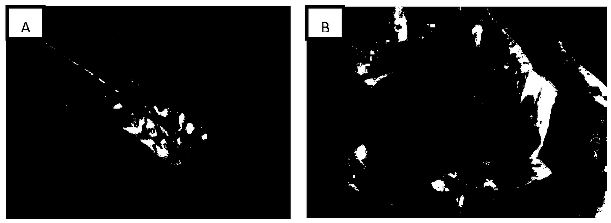
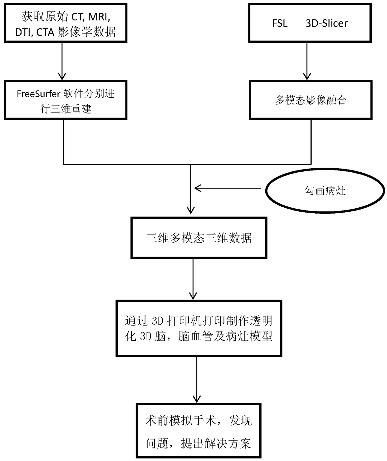
[0044] Step 2. Process the imaging data to realize the reconstruction of the brain and cerebrovascular; MRI is better for brain tissue display, a...


PUM


Abstract

The invention provides a cerebral cortex structure, a cerebral blood vessel, an intracerebral lesion 3D printing model and a preparation method thereof. The preparation method comprises the followingsteps that 1, multiple types of original image data of the brain of a patient are obtained and output; 2, three-dimensional reconstruction of the cerebral cortex, white matter fiber tracts and the blood vessel is conducted at least through three-dimensional reconstruction software according to multiple types of output original image data, and a lesion is sketched out' 3, multimode image fusion isconducted on reconstructed models by utilizing image analysis software to obtain three-dimensional multimode data; 4, based on the obtained three-dimensional multimode data, 3D printing is performed according to the proportion 1:1 of the data to a photosensitive resin material, and a transparent 3D printing model including the cerebral cortex structure, the cerebral blood vessel and the intracerebral lesion is obtained.

Description

technical field [0001] The invention relates to a method for preparing a 3D printing model of cerebral cortex structure, cerebrovascular and intracerebral lesions, which can be used as an auxiliary surgical system to simulate surgical operations before surgery, and belongs to the medical field. Background technique [0002] Neurosurgery is one of the most risky and difficult professions in clinical medicine, with high morbidity and mortality rates, and precise targeting is required in the diagnosis and treatment process; and the scope of neurosurgery is limited, requiring the operator to be familiar with Local anatomical levels, and the establishment of a three-dimensional concept. Establishing a three-dimensional concept from a two-dimensional image in the mind is a process in which the comprehensive strength of neurosurgeons is greatly improved. The neuronavigation system simulates the digital images of computed tomography (CT) and magnetic resonance imaging (MRI). It est...

Claims


Application Information

Patent Timeline
IPC IPC(8): A61B34/10A61B34/20B33Y80/00
CPCA61B34/10A61B34/20A61B2034/101A61B2034/105A61B2034/107A61B2034/108A61B2034/2065B33Y80/00
Inventor 徐纪文王昌泉周洪语叶晓来赵晨杰马军峰刘强强
Owner RENJI HOSPITAL AFFILIATED TO SHANGHAI JIAO TONG UNIV SCHOOL OF MEDICINE
Why Eureka
Try Eureka