Auto-focus methods and systems for digital imaging using mutli-spectral trajectories

A technology of digital imaging system and scanning trajectory, which is applied in the parts, optics, image communication and other directions of the TV system, which can solve the problems of large data file size and long scanning time, etc.

Active Publication Date: 2018-09-28
VENTANA MEDICAL SYST INC
View PDF6 Cites 0 Cited by
  • Summary
  • Abstract
  • Description
  • Claims
  • Application Information

AI Technical Summary

Problems solved by technology

This approach provides high image quality while resulting in excessively long scan times and large data file sizes

Method used


Image

  • Auto-focus methods and systems for digital imaging using mutli-spectral trajectories
  • Auto-focus methods and systems for digital imaging using mutli-spectral trajectories
  • Auto-focus methods and systems for digital imaging using mutli-spectral trajectories

Examples


Embodiment Construction

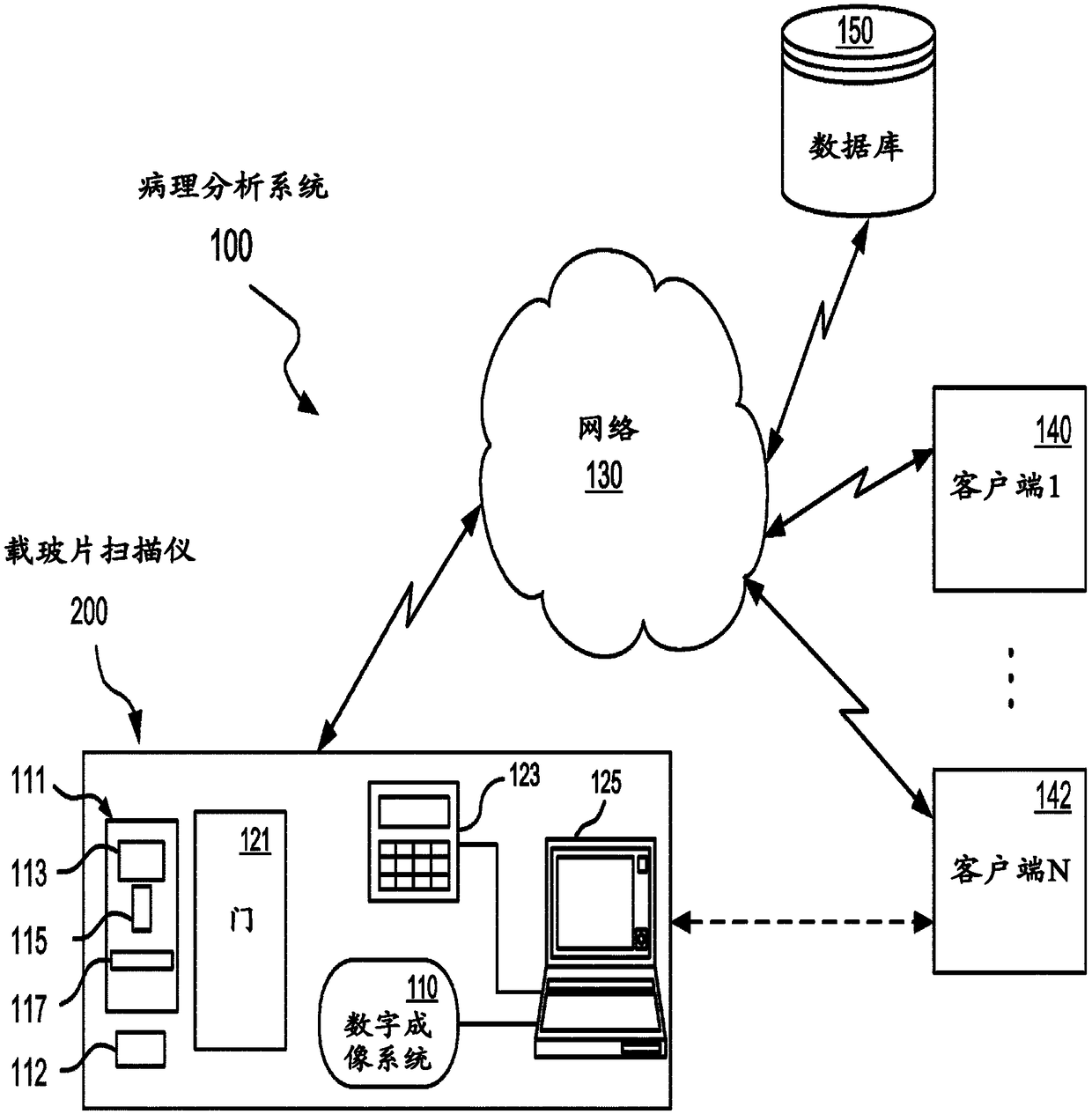
[0045] figure 1 A pathology analysis system 100 according to one embodiment of the present application is shown operating in a network environment for implementing an autofocus technique for digital imaging using multispectral trajectories (also referred to herein as "automated slide scanning" or "automated slide scanning" scanning"). As used herein, "autofocus" refers to the automatic digitization of microscope slides such that the resulting images are useful for pathologists to make a diagnosis.

[0046] Microscope slides are also referred to herein as "specimen slides" or "glass slides". The microscope slides of the present application are intended to hold very thin layers of biological samples (eg, cells or tissue). Samples are typically stained or otherwise labeled for any one or more of the following functions:

[0047] (i) absorption of light transmitted through the slide ("brightfield imaging"),

[0048] (ii) scatters light from an external source, or

[0049] (i...


PUM


Abstract

A method and associated method and computer program product for acquiring focused images of a specimen on a slide, by determining optimal scanning trajectories. The method includes capturing a relatively low magnification image of the slide to locate the specimen, forming a grid that includes an arrangement of grid points, overlaying at least part of the grid over a field of view that covers at least part of the specimen, capturing a relatively high magnification Z-stack of images of the specimen within the field of view, determining a best focus for each grid point within said at least part of the grid to form a resulting grid of three dimensional points, and based on the resulting grid, determining one or more three dimensional scanning trajectories.

Description

[0001] related application [0002] This application claims priority to US Patent Application No. 62 / 268,165, filed December 16, 2015, which is hereby incorporated by reference in its entirety. [0003] related application [0004] The present application relates generally to computer-based autofocus systems and related methods for imaging microscope slides. More specifically, the application relates to automated slide scanning using multi-spectral trajectories to obtain high quality image data at high slide digitization rates. Background technique [0005] Digital pathology equipment is commonly used to produce digital images of microscope slides. Pathologists and histologists typically visually inspect digital images to gain information about tissue samples and identify the most appropriate treatments to improve clinical outcomes. To produce color images, automated slide scanners typically acquire images in the red, green and blue channels separately and combine these i...

Claims


Application Information

Patent Timeline
IPC IPC(8): G02B7/38G02B21/36H04N1/387
CPCH04N1/3876G02B21/367H04N23/67G02B21/006
Inventor J·布里登J·马丁A·萨卡尔
Owner VENTANA MEDICAL SYST INC
Why Eureka
Try Eureka