

Application of miRNA (micro-ribonucleic acid) miR-21 to glaucoma treatment
A technology of mirna-21 and .mirna-21 is applied in the application field of small molecule nucleic acid miR-21 in the treatment of glaucoma, which can solve the problem of common adverse reactions in the eyes of patients.
- Summary
- Abstract
- Description
- Claims
- Application Information
AI Technical Summary
Problems solved by technology
Method used
Image

Examples
Embodiment 1
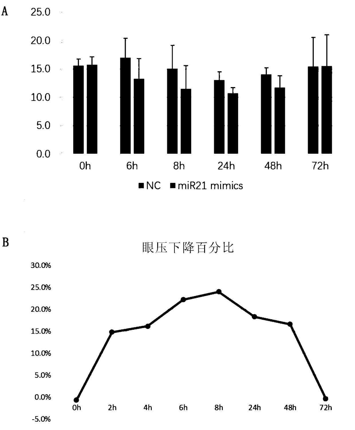
[0118] Effect of miR-21 on intraocular pressure
[0119] Combining miR-21mimics with transfection reagents in vivo, the drug was injected into the anterior chamber of normal mice by anterior chamber injection, and a negative control (Negative Control, NC) was given to the contralateral eye (the negative control was a nonsense random sequence ). Administration of mouse anterior chamber under microscope figure 1 shown.
[0120] Changes in intraocular pressure were measured with a tonometer before and after administration. When it was confirmed that the change in intraocular pressure was the most obvious, the mouse eyeball was completely removed, and the outflow rate of aqueous humor was measured using a mouse isolated eyeball perfusion model. The specific operation method is as follows:
[0121] Use a 33G needle to puncture the anterior chamber, and maintain constant pressure (12mmHg) perfusion for 30 minutes, and then measure the changes in the outflow and outflow rate of n...
Embodiment 2
[0125] Effect of miR-21 on cell permeability
[0126] Using porcine angular aqueous plexus (AAP) cells to simulate the Schlemm duct cells in the human trabecular meshwork pathway, using virus transfection to up-regulate the amount of intracellular miR-21, and measuring the transendothelial electric resistance (TEER) of the cells , according to the inverse ratio of transmembrane resistance to cell permeability, reflecting the resistance in the outflow pathway of aqueous humor.
[0127] The result is as Figure 5 Shown: Permeability increased after cells were transfected with viruses overexpressing miR-21mimics. 5A and 5B overexpressed miR-21mimics by virus transfection, and the transfection efficiency was 70%. Compared with transfection of empty virus (Negative Control group), miR-21mimics transmembrane cell resistance of 5C was significantly reduced. That is, up-regulation of miR-21mimics can increase the permeability of porcine Schlemm duct endothelial cells (p<0.01), and r...
Embodiment 3
[0129] miR-21 targets the trabecular pathway to achieve multi-target blood pressure reduction
[0130] Obtaining chamber angle tissue: The mice transfected for 24 hours were killed by cervical dislocation, and the eyeballs were taken out immediately. Use ophthalmic micro-scissors to cut along the equator of the eyeball, take out the crystal, gently pull out the iris with micro-tweezers, take out the root of the iris as much as possible, carefully cut off the cornea along the limbus, cut off the posterior structure about 0.5mm behind the limbus, and take the cornea The structure between the edge and the back is the angle tissue. Histologically, it contains trabecular meshwork, Schlemm's canal, and to a lesser extent the cornea, iris root, and external sclera.
[0131] Protein pathway verification: fresh chamber angle tissues transfected with miR-21mimics and NC were taken, lysed with RIPA lysate on ice for 1 hour, and ultrasonically shattered. After the protein concentration ...
PUM
 Abstract
Description
Claims
Application Information