Radiation pneumonitis CT (Computed Tomography) quantitative detection method used for radiobiology experiment

A quantitative detection method, technology of radiation pneumonitis, applied in measuring devices, scientific instruments, material analysis using wave/particle radiation, etc., can solve the problems of combined cardiotoxicity, affecting the accuracy of results, affecting the accuracy and reliability of models, etc. , to achieve the effect of increasing accuracy, reducing dependence, reducing experimental cost and effort

Inactive Publication Date: 2018-12-11
ZHEJIANG CANCER HOSPITAL
View PDF0 Cites 3 Cited by
  • Summary
  • Abstract
  • Description
  • Claims
  • Application Information

AI Technical Summary

Problems solved by technology

However, there are still many technical deficiencies in these detection and evaluation methods.
[0004] First, physiological parameters (i.e., respiratory rate, lung compliance, lethality, or body weight) can more objectively reflect abnormal gas exchange due to pulmonary restrictive ventilation, diffuse interstitial inflammation, or scarring, but these parameters are susceptible to many other factors. Interference of factors, such as temperature, infection, combined cardiotoxicity, etc.
[0005] Second, when using low-dose irradiation, only a small amount of pneumonia can be observed in lung tissue sections. These physiological indicators cannot truly reflect lung lesions in the milder lesions or in the self-healing stage: no obvious changes in respiratory rate, Individual death or weight loss (such as X-ray dose <10.5Gy), prone to false negatives
However, in this false negative situation, in mathematical modeling, the measured value of gradient dose (from low to high) is usually required for calculation and simulation, that is, the sensitivity of the detection value of the low dose group will directly affect the accuracy and reliability of the model
[0006] Third, due to the greater compensatory capacity of lung function, for the evaluation of early lung injury (ie, the inflammatory stage), histological evidence of lung interstitial thickening, inflammatory cell proliferation or infiltration is associated with individual death or increased respiratory rate in mice. There is no direct correlation, which affects the accuracy of the results to a certain extent
However, due to the small size of individual mice (the transverse diameter of the lungs of adult mice is only about 1-1.5 cm) and the resolution of CT scanning is limited, even high-resolution or professional micro-CT images cannot accurately diagnose radiation pneumonitis in mice
In view of the above-mentioned defects in the current experiments on radiation-induced lung injury mice, which cannot achieve the purpose of objective and accurate quantitative evaluation, the detection method of the present invention adopts computer-assisted imaging diagnosis (computer-assisted radiology) technology to realize a simple, accurate and quantitative evaluation of pneumonia. developmental approach

Method used


Image

  • Radiation pneumonitis CT (Computed Tomography) quantitative detection method used for radiobiology experiment
  • Radiation pneumonitis CT (Computed Tomography) quantitative detection method used for radiobiology experiment
  • Radiation pneumonitis CT (Computed Tomography) quantitative detection method used for radiobiology experiment

Examples


Embodiment 1

[0035] Embodiment 1 radiation biology (mice) experiment uses radiation pneumonitis CT quantitative detection method

[0036]A conventional medical linear accelerator was used to irradiate the whole lung field of mice to complete the modeling of radiation pneumonitis in mice. Mice were under anesthesia and well fixed.

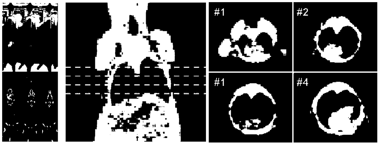
[0037] refer to figure 1 At different time points after the whole lung field of mice was irradiated, images were collected using conventional medical computed tomography (CT) equipment with low-dose scanning parameters; followed by 3D image reconstruction and 3D lung segmentation based on automatic region growing.

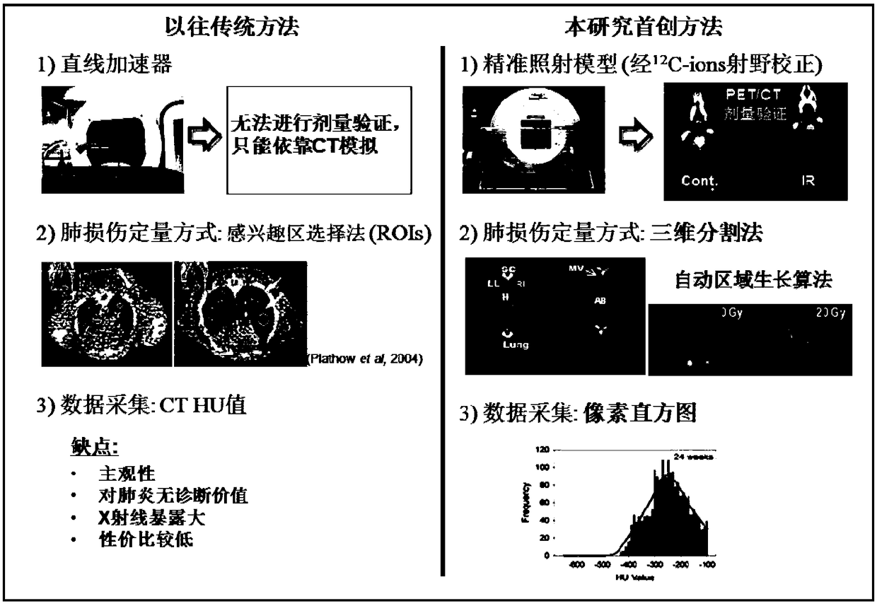
[0038] refer to figure 2 , an intuitive comparison between the CT non-invasive quantitative detection technology of the present invention and the traditional method. In the past, traditional CT image evaluation usually used Regions of interests (ROIs), its main disadvantages include: high subjectivity (relying on the experimenter’s subjective out...

Embodiment 2

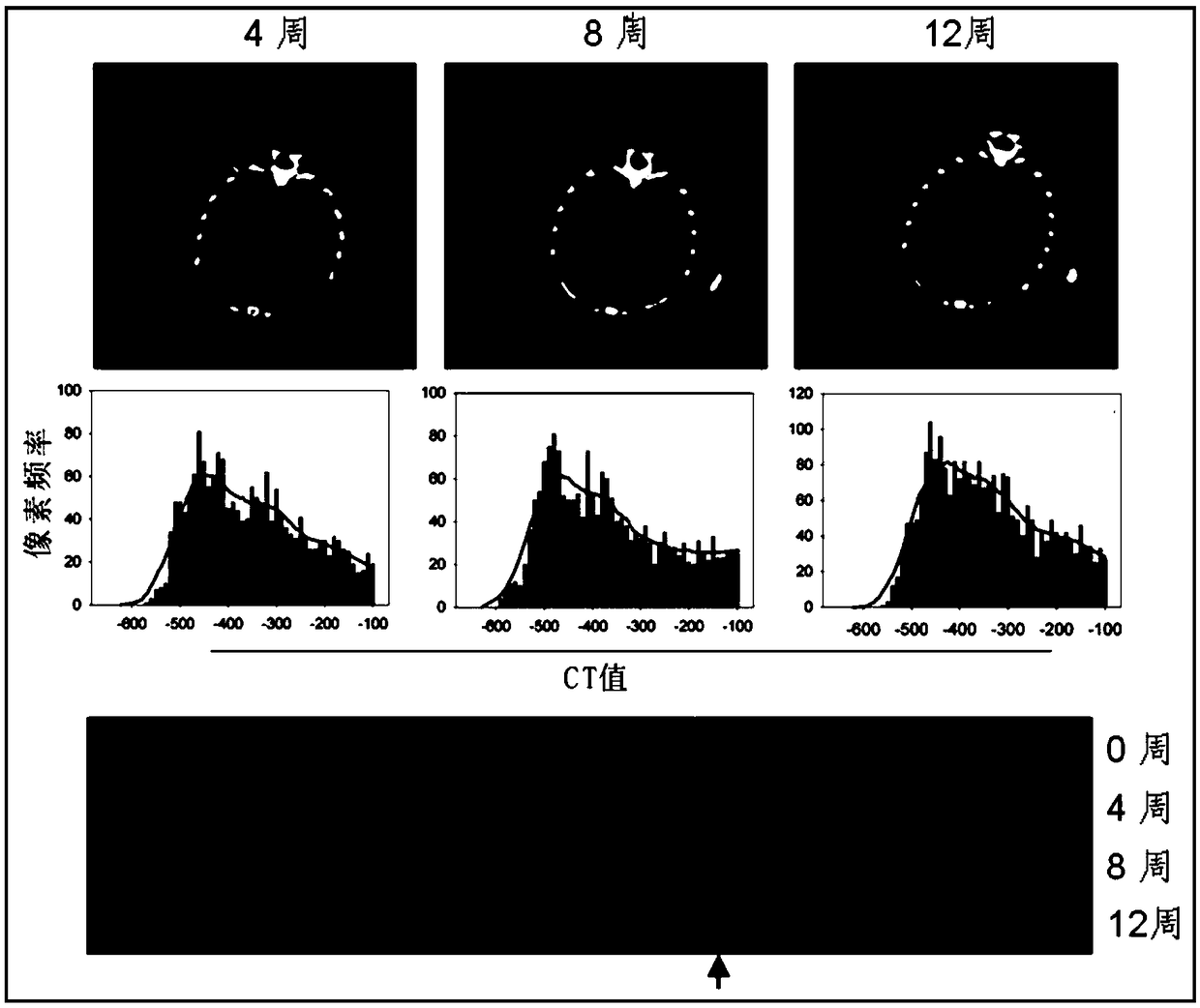
[0043] Example 2: Quantitative detection of pneumonia was carried out 12 weeks after C57BL6 at the age of 8-10 weeks received whole lung field irradiation. The specific methods are as follows:

[0044] 1. Irradiation and grouping of animal experiments: C57BL6 female mice aged 8-10 weeks were used and raised at SPF level. Light for 12h, free access to food and water.

[0045] 2. After induction of anesthesia by isoflurane before irradiation, transfer to a dedicated mouse chest irradiation device and fix it properly. The whole lung field was irradiated by conventional medical linear accelerator.

[0046] 3. Give a single dose of 20Gy irradiation.

[0047] 4. The blank control group did not receive X-ray irradiation (0Gy). 12 mice were randomly assigned to each group of the control group and each dose of the experimental group;

[0048] 5. CT scans were performed every 4 weeks before and after irradiation (time points of 0, 4, 8, and 12 weeks respectively); low-dose scanning ...


PUM


Abstract

The invention discloses a radiation pneumonitis CT (Computed Tomography) quantitative detection method used for a radiobiology experiment. Through a CT low-dosage scanning parameter, a scanning imageis subjected to three-dimensional reconstruction, then, a CT three-dimensional segmentation image pixel histogram is obtained on the basis of an automatic region growth lung image matrix three-dimensional segmentation technology, a pneumonitis index algorithm is adopted through a pneumonitis degree quantitative criterion, and a mouse pneumonitis index is obtained to finish noninvasive quantitativedetection. The method effectively appears in noninvasive quantitative detection methods which aim at the mouse radiation pneumonitis model at home and abroad for the first time, and is an experimentdetection method which can objectively and accurately quantify the generation and the development of the mouse pneumonitis in real time, and the accuracy, the simplification, the stability and the efficiency of radiation inducibility mouse pneumonitis detection can be obviously improved. While experiment cost and vigor can be greatly reduced, the dependency of small animal minitype CT is reduced,experience cost is lowered, and the method has the obvious advantages of no trauma, high speed in scanning, high accuracy, good repeatability and the like.

Description

technical field [0001] The technology of the present invention belongs to experimental methods, relates to a CT quantitative detection method, in particular to a CT quantitative detection method for radiobiology (mice) experiments of radiation pneumonitis. Background technique [0002] The mouse model of radiation pneumonitis is an important tool for studying the mechanism of normal tissue radiation response, respiratory disease and immune response, and plays an irreplaceable role in the research and development of radiation biology, radiotherapy and anti-lung injury drugs. In the early stage of radiation lung injury, the main manifestation is radiation pneumonitis (usually 1-3 months after secondary radiation), due to the activation of a large number of T lymphocytes to produce an immune response, a type of lymphocytic alveolitis; in the late stage, it can develop into interstitial lung fibrosis (about 6 months after irradiation). Once pulmonary fibrosis occurs, the conseq...

Claims


Application Information

Patent Timeline
Patent Type & Authority Applications(China)
IPC IPC(8): G01N23/046
CPCG01N23/046
Inventor 周成张鹏陈明
Owner ZHEJIANG CANCER HOSPITAL
Why Eureka
Try Eureka