

Construction method and application of retinal neovascular disease model
A new blood vessel and disease model technology, applied in the biological field, can solve the problems of lack of animal models and achieve the effect of broad application prospects
- Summary
- Abstract
- Description
- Claims
- Application Information
AI Technical Summary
Problems solved by technology
Method used
Image

Examples
Embodiment 1
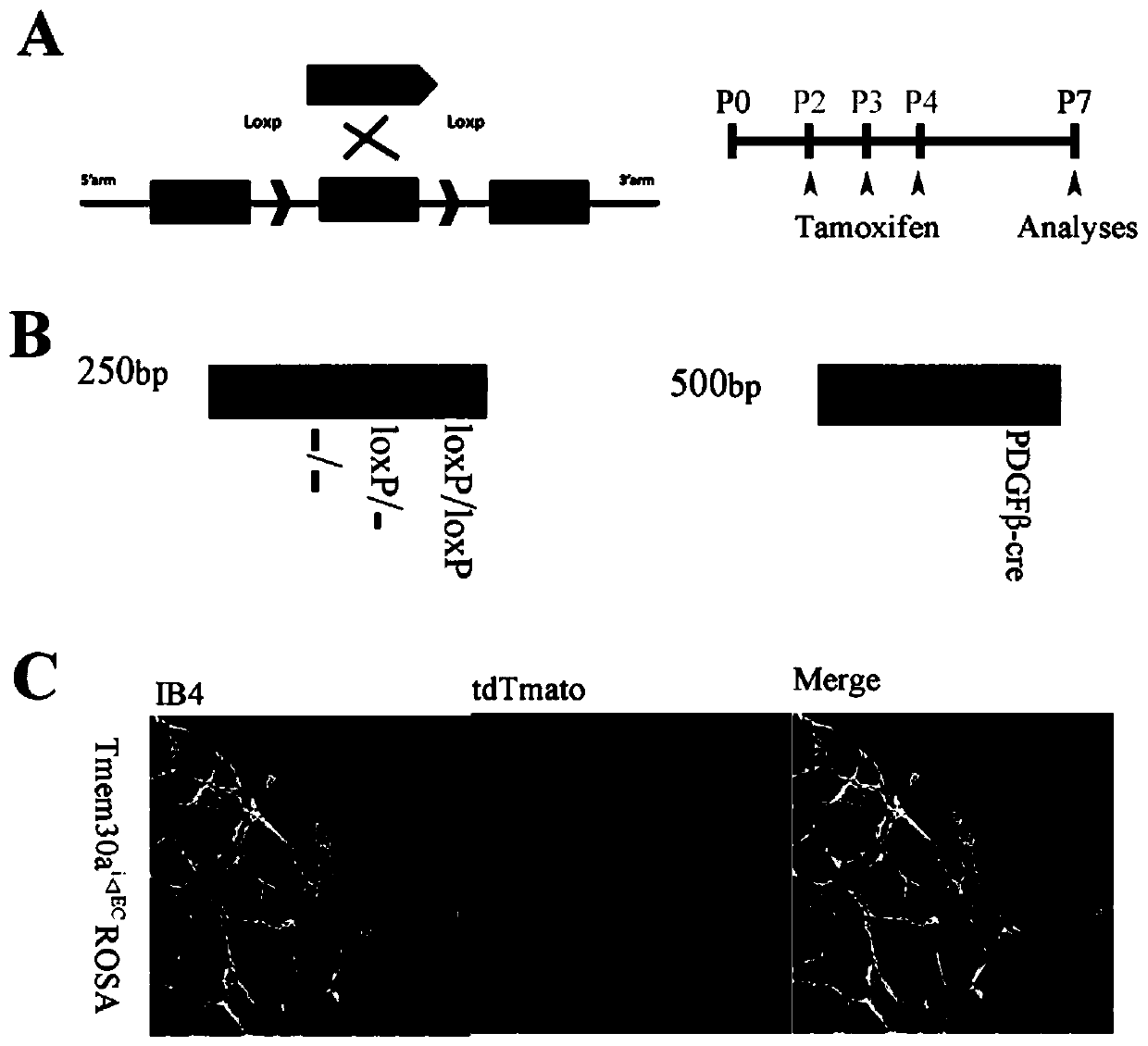
[0057] In this example, mice are used as target animals, and the method for constructing the retinal neovascular disease model provided by the present invention is described. The route of knocking out the TMEM30A gene is as follows: figure 2 As shown in A, the specific operation is as follows:
[0058] 1. Refer to the method of the Chinese patent application No. 2017103803265, named the construction method and application of the mouse model of pancreatic beta cell conditional knockout of Tmem30a gene, to obtain the conditional knockout of Tmem30a gene (exon No. 3) homozygous small mouse;
[0059] 2. The Tmem30a gene conditional knockout homozygous mice were mated with PDGFβ-Cre ER transgenic mice to obtain Tmem30a gene conditional knockout mice for vascular endothelial cells.
[0060] When PDGFβ-Cre ER transgenic mice were crossed with Tmem30a knockout homozygous mice, half of the offspring carried both PDGFβ-Cre ER and Tmem30a conditional knockout. This animal was mated wi...
Embodiment 2
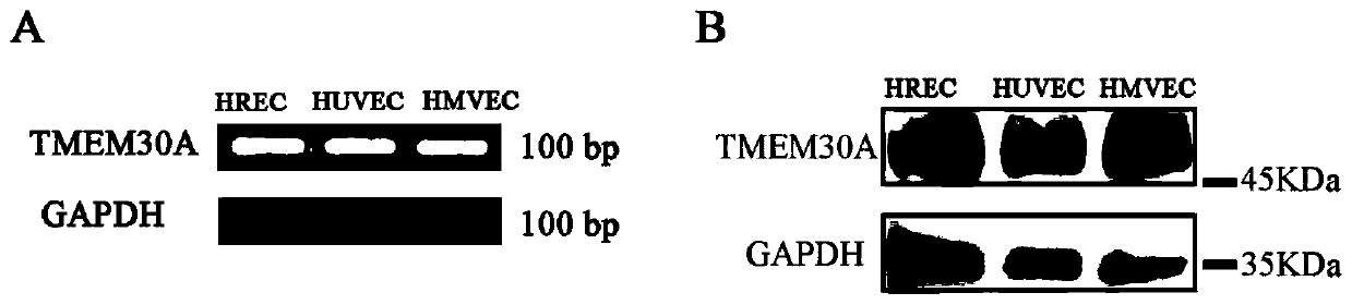
[0062] 1 RT-PCR method was used to detect the expression of TMEM30A gene in three kinds of vascular endothelial cells.
[0063] Methods: The total RNA of three kinds of vascular endothelial cells human retinal vascular endothelial cells (HREC), human umbilical vein vascular endothelial cells (HUVEC) and human microvascular endothelial cells (HMVEC) were extracted respectively, and then cDNA synthesis kit (Invitrogen, Waltham, MA, USA) to synthesize cDNA. Primers were designed according to the cDNA sequence of TMEM30A:
[0064] TMEM30A-H-F: 5'-TCGGTCTCATCTTCATTCCC-3';
[0065] TMEM30A-H-R: 5'-GGAAGGCTCTGTTCCGGTAT-3'.
[0066] Using the extracted cDNA as a template, RT-PCR was performed. After amplification, electrophoresis was carried out, and the results were shown in figure 1 .
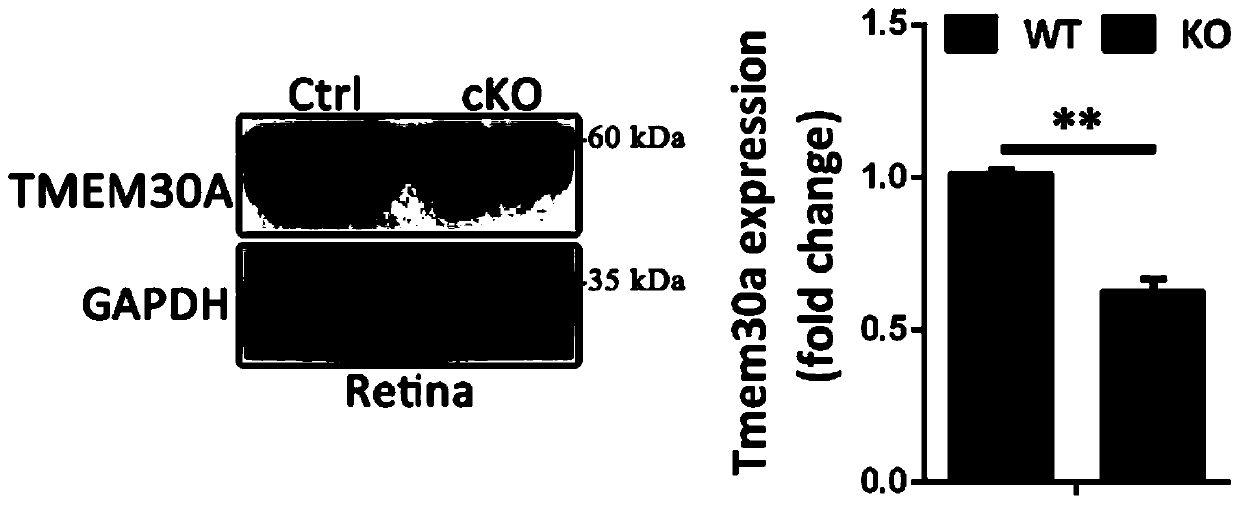
[0067] see results figure 1 A, it can be seen that the expression of TMEM30A gene in human retinal vascular endothelial cells (HREC), human umbilical vein vascular endothelial cells (HUVEC) and...
Embodiment 3
[0081] Identification of the genotype of the mouse that specifically knocked out the Tmem30a gene in the vascular endothelial cells in Example 1.
[0082] method:
[0083] (1) Cut a little tissue sample from the mouse tail and place it in a clean 1.5ml centrifuge tube;
[0084] (2) Add 100μl of lysate (40mM NaOH, 0.2mM EDTA solution) to the centrifuge tube, and heat in a metal bath at 100°C for 1h; (3) Take out the centrifuge tube, cool to room temperature, and add 100μl of neutralizing solution (40mM Tris-HCl, pH 5.5), centrifuged at 10000 g for 2 min, and the supernatant was taken for mouse genotype identification.
[0085] (4) PCR amplification: configure the PCR reaction system according to the following system
[0086]
[0087] The primer sequences are as follows:
[0088] Tmem30a-Forward sequence: 5'-ATTCCCCTTCAAGATAGCTAC-3';
[0089] Tmem30a-Reverse sequence: 5'-AATGATCAACTGTAATTCCCC-3';
[0090] PDGFβ-Forward sequence: 5'-GCCGCCGGGATCACTCTCG-3';
[0091] PDGFβ...
PUM
| Property | Measurement | Unit |
|---|---|---|
| thickness | aaaaa | aaaaa |
Abstract
Description
Claims
Application Information