Fluorescence microscopic imaging device and method by using delayed fluorescence

A delayed fluorescence and microscopic imaging technology, which is applied in the field of fluorescence imaging, can solve the problems of high price, large device volume, and precise and complex structure, and achieve the effects of simplifying system complexity, improving portability, and saving costs

Active Publication Date: 2019-05-31
NANJING UNIV
View PDF6 Cites 3 Cited by
  • Summary
  • Abstract
  • Description
  • Claims
  • Application Information

AI Technical Summary

Problems solved by technology

However, the traditional fluorescence microscopy device based on the lens system not only has a small volume of one-time observable sample, but also has a larg

Method used


Image

  • Fluorescence microscopic imaging device and method by using delayed fluorescence
  • Fluorescence microscopic imaging device and method by using delayed fluorescence
  • Fluorescence microscopic imaging device and method by using delayed fluorescence

Examples


Example Embodiment

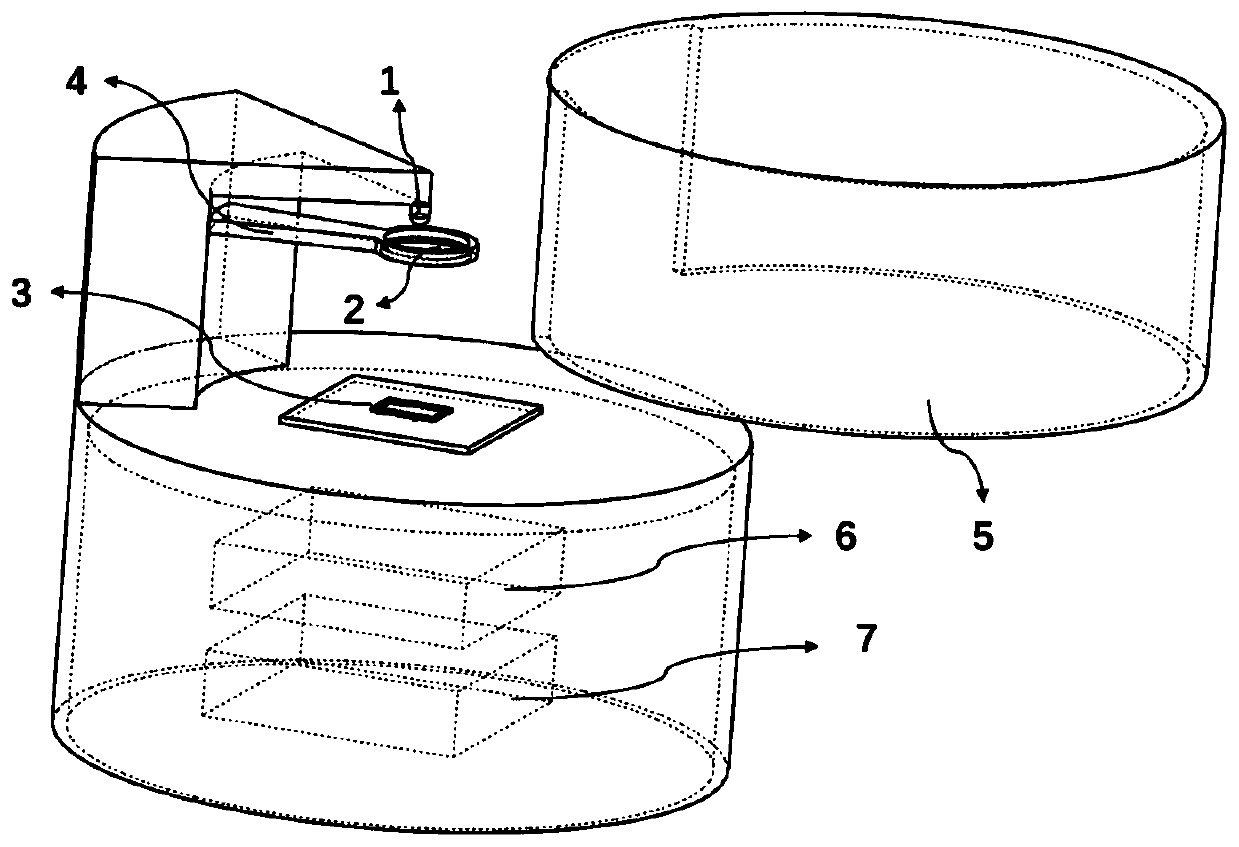
[0023] An embodiment of the present invention provides a fluorescence microscope imaging device using delayed fluorescence, and its overall structure is as follows figure 1 As shown, it includes an excitation light source module, a microscopic imaging module, a timing control module 6 and an image data processing and display module 7.

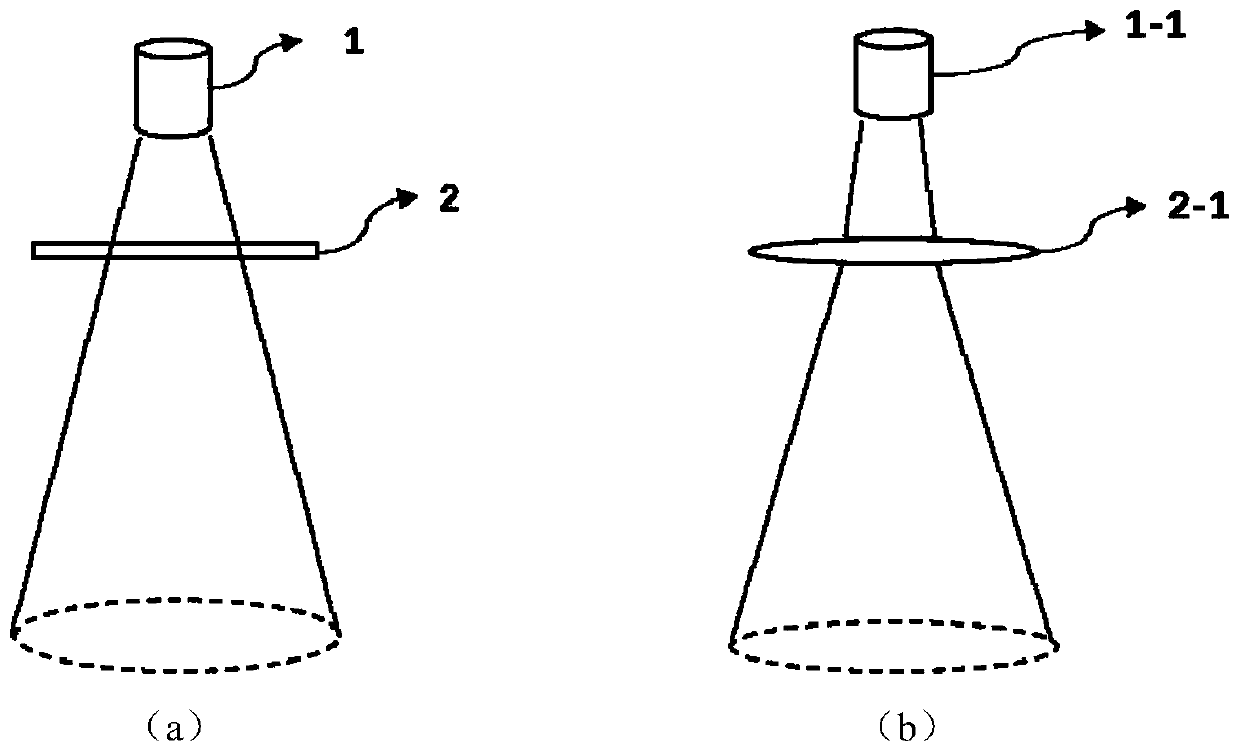
[0024] The excitation light source module is used to generate excitation light in a specific wavelength range to excite the delayed fluorescence signal in the delayed fluorescence sample. The module is placed directly above the fluorescence microscope imaging device, and the light-emitting surface covers the entire photosensitive area of ​​the image sensor chip 3. There are many options for the excitation light source module, which can be composed of LED light source 1 combined with band pass filter 2, such as figure 2 As shown in (a), it can also be composed of a specific wavelength laser diode 1-1 combined lens 2-1 as figure 2 As shown in (b), ...


PUM

PropertyMeasurementUnit
Thicknessaaaaaaaaaa

Abstract

The present invention proposes a fluorescence microscopic imaging device and method by using delayed fluorescence. The prepared delayed fluorescent sample is directly added to a sample microcavity atthe surface of an image sensor, the fluorescence intensity can be maintained for a period of time after the excitation light source is extinguished by using the delayed fluorescence self-characteristic, a working mode of the image sensor is arranged to collect fluorescence signals after the excitation light source is extinguished so as to achieve the fluorescence signal detection. The fluorescencemicroscopic device does not need a traditional imaging lens system and does not an optical filter for filter of excitation light so as to greatly save the cost, simplify the system complexity, improve the convenience of the system, achieve the statistical observation of large field of view and large volume samples and provide some reference and convenience for biomedical analysis, drug screeningand cell processing in certain scene.

Description

technical field [0001] The invention relates to the field of fluorescence imaging, in particular to a lensless fluorescence microscopic imaging device and a method thereof. Background technique [0002] Fluorescence microscopy has been widely used in biomedical analysis, drug screening, cell processing and detection, and has become an essential tool in the field of life science research. However, traditional fluorescence microscopy devices show weakness and insufficiency in some research scenarios. The characterization and detection of rare cells (such as circulating tumor cells) is an important topic in the field of biomedicine, because some rare cells play a vital role in disease diagnosis, but the concentration of these cells is often very low, and some even have only Therefore, in order to perform accurate monitoring and analysis, it is necessary to observe a sufficient volume of samples to obtain a sufficient number of target cells. However, the traditional fluorescen...

Claims


Application Information

Patent Timeline
IPC IPC(8): G01N21/64
Inventor 闫锋曹旭杨程岳涛孟云龙马浩文卜晓峰林岚昆沈心雨
Owner NANJING UNIV
Why Eureka
Try Eureka