A method for processing immunohistochemical images
A technology of immunohistochemistry and image processing, which is applied in image data processing, image analysis, instruments, etc., can solve the problems of not clear enough images, fading, and the influence of image processing results, and achieve clear images, easy identification, and prolong the fading time. Effect
- Summary
- Abstract
- Description
- Claims
- Application Information
AI Technical Summary
Problems solved by technology
Method used
Image
Examples
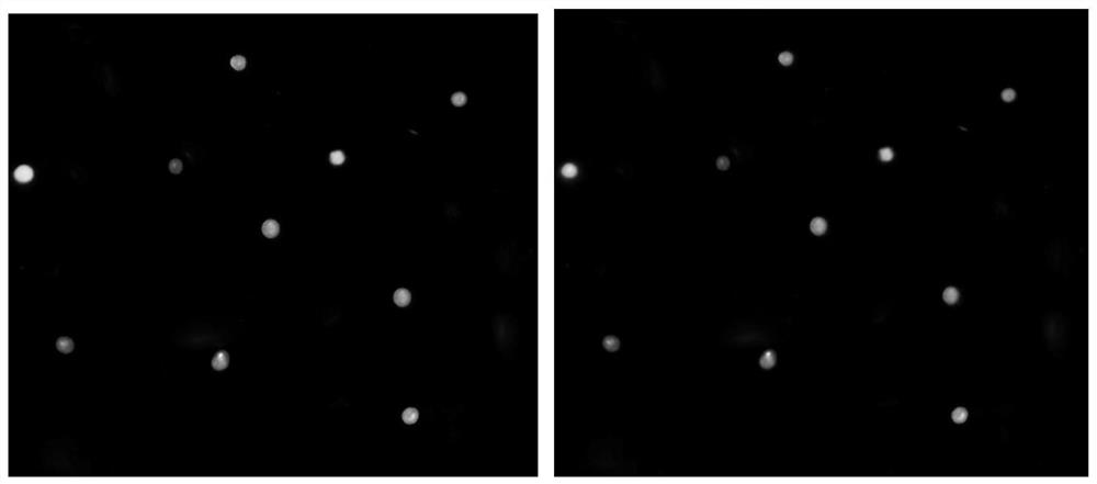
Embodiment 1
[0028] A method for processing immunohistochemical images, comprising the following steps:
[0029] (a) Perform full-stained immunohistochemical staining on tissue sections to obtain digital immunohistochemical images; wherein, the staining process is: after mixing DAB staining solution with potassium dihydrogen phosphate, gelatin, and acetic acid (mass ratio: 100:0.2 :0.03:0.06) to stain the positive product, rinse with sodium dihydrogen phosphate solution (concentration of sodium dihydrogen phosphate solution is 0.2g / L) for 2 to 3 minutes, rinse with water, and mix hematoxylin staining solution with glycerol and betaine (Mass ratio: 100:0.1:0.03) After counterstaining cell nuclei, hydrochloric acid alcohol differentiation, water washing, dehydration, transparent, mounting, microscopic examination;
[0030] (b) convert the immunohistochemical image described in (a) step from RGB color space to YIQ color space, and normalize the value of the Y component of the transformed imag...
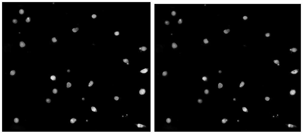
Embodiment 2
[0040] A method for processing immunohistochemical images, comprising the following steps:
[0041] (a) Perform full-staining immunohistochemical staining on tissue sections to obtain digital immunohistochemical images; wherein, the staining process is: mixing DAB staining solution with potassium dihydrogen phosphate, gelatin, and acetic acid (mass ratio: 100:0.1: 0.02:0.05) to stain the positive product, wash with sodium dihydrogen phosphate solution (the concentration of sodium dihydrogen phosphate solution is 0.2g / L) for 2 to 3 minutes, rinse with water, and mix hematoxylin staining solution with glycerol and betaine (Mass ratio: 100:0.2:0.05) After counterstaining cell nuclei, hydrochloric acid alcohol differentiation, water washing, dehydration, transparent, mounting, microscopic examination;
[0042] (b) convert the immunohistochemical image described in (a) step from RGB color space to YIQ color space, and normalize the value of the Y component of the transformed image ...
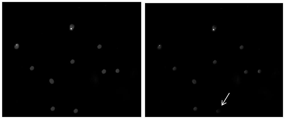
Embodiment 3
[0051] A method for processing immunohistochemical images, comprising the following steps:
[0052] (a) Perform full-staining immunohistochemical staining on tissue sections to obtain digital immunohistochemical images; wherein, the staining process is: mixing DAB staining solution with potassium dihydrogen phosphate, gelatin, and acetic acid (mass ratio: 100:0.2: 0.05:0.1) After staining the positive product, rinse with sodium dihydrogen phosphate solution (concentration of sodium dihydrogen phosphate solution is 0.3g / L) for 2 to 3 minutes, rinse with water, and mix hematoxylin staining solution with glycerol and betaine (Mass ratio: 100:0.2:0.05) After counterstaining cell nuclei, hydrochloric acid alcohol differentiation, water washing, dehydration, transparent, mounting, microscopic examination;
[0053] (b) convert the immunohistochemical image described in (a) step from RGB color space to YIQ color space, and normalize the value of the Y component of the transformed imag...
PUM
 Abstract
Description
Claims
Application Information
 - R&D
- Intellectual Property
- Life Sciences
- Materials
- Tech Scout
- Unparalleled Data Quality
- Higher Quality Content
- 60% Fewer Hallucinations
Browse by: Latest US Patents, China's latest patents, Technical Efficacy Thesaurus, Application Domain, Technology Topic, Popular Technical Reports.
© 2025 PatSnap. All rights reserved.Legal|Privacy policy|Modern Slavery Act Transparency Statement|Sitemap|About US| Contact US: help@patsnap.com


