
Holographic positive electron concentration imaging method and system

An imaging method and imaging system technology, applied in the field of medical particle physics applications, can solve problems such as insufficient improvement of imaging key performance and focus on spatial resolution, so as to solve the aggravation of statistical noise, improve spatial resolution and sensitivity, and reduce background noise. Effect

Inactive Publication Date: 2019-07-12
NANCHANG UNIV
View PDF0 Cites 0 Cited by
  • Summary
  • Abstract
  • Description
  • Claims
  • Application Information

AI Technical Summary

Problems solved by technology

The completely fixed general reconstruction method and the idea of ​​focusing too much on spatial resolution make the gamma mode PET reconstruction method always face the embarrassment of insufficient improvement in key imaging performance in practical applications

Method used


Image

  • Holographic positive electron concentration imaging method and system
  • Holographic positive electron concentration imaging method and system
  • Holographic positive electron concentration imaging method and system

Examples


Embodiment Construction

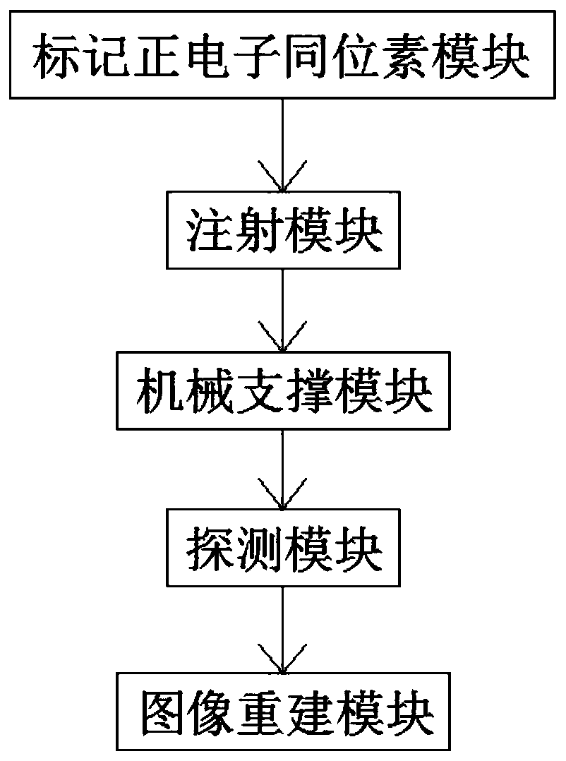
[0035] Embodiments of the present invention will be further described below in conjunction with the accompanying drawings.

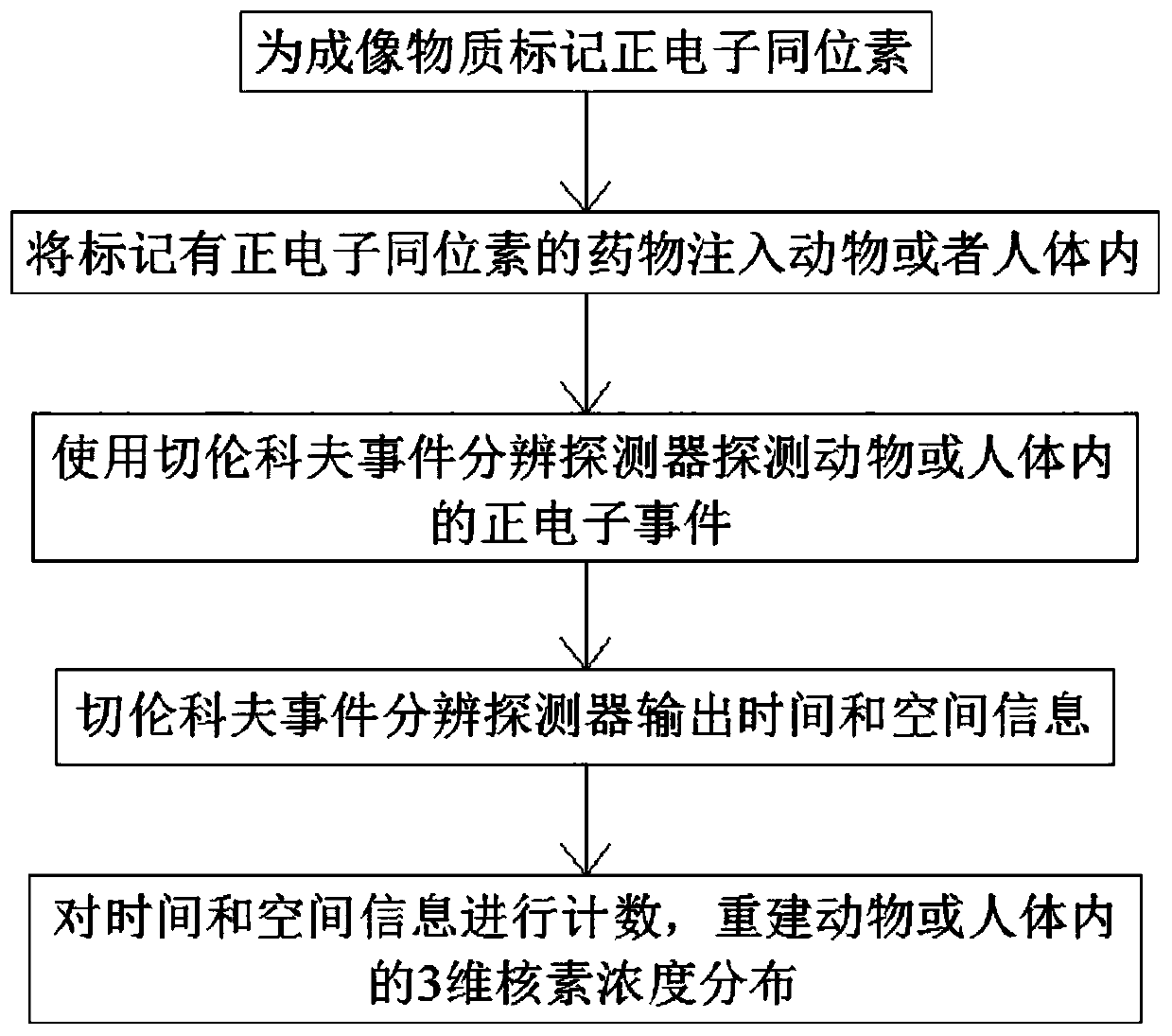
[0036] Such as Figure 1-2 As shown, a holographic positron concentration imaging method comprises the following steps:

[0037] Step 1: labeling positron isotopes for imaging substances;

[0038]Step 2: Inject the drug labeled with the positron isotope into the animal or human body so that it is distributed in the animal or human body;

[0039] Step 3: Detect positron events in animals or humans using Cerenkov event-resolving detectors;

[0040] Step 4: Cherenkov event resolution detector outputs time and space information;

[0041] Step 5: Count the time and space information of the Cerenkov event resolution detectors, and reconstruct the 3-dimensional nuclide concentration distribution in animals or humans.

[0042] The positron isotope in said step 1 refers to the proton-rich isotope produced by the medical cyclotron, such as commonly used radioa...


PUM


Abstract

The invention discloses a holographic positive electron concentration imaging method. The method comprises the steps of labeling a positive electron isotope for an imaging substance; injecting a druglabeled with the positive electron isotope into an animal or the human body, so that the drug is distributed in the animal or the human body; utilizing a Cherenkov event resolution detector for detecting a positive electron event in the animal or the human body; utilizing the Cherenkov event resolution detector for outputting time and space information; counting the time and space information of the Cherenkov event resolution detector, and reconstructing three-dimensional nuclide concentration distribution in the animal or the human body. The method can effectively improve the spatial resolution and sensitivity of the gamma mode PET, is particularly suitable for clinical PET which has a very high requirement for the accuracy and precision, and is widely applied in cancers and cardiovascular diseases. The high spatial resolution and time resolution facilitate improvement of the imaging quality; the high sensitivity facilitates lowering of imaging background noise, and the problem that statistical noise influence is intensified under the high resolution is solved.

Description

technical field [0001] The invention belongs to the technical field of medical particle physics application, and in particular relates to a holographic positron concentration imaging method and system. Background technique [0002] During the past decade, research on gamma-mode PET has focused on improvements in spatial resolution and sensitivity. The YAP-PET developed in Italy uses a PET / SPECT dual-mode structure, and the size of a single YAP scintillation crystal is 2×2×30mm 3 , can provide a reconstruction image spatial resolution of 1.8mm FWHM. The MicroPET Focus 120 of Washington University is in lutetium orthosilicate (Lu 2 SiO 5 , LSO for short) scintillation crystal and position-sensitive photomultiplier tube (Position-Sensitive Photomultiplier, PSPMT for short) are coupled by optical fiber. At the center point of the Field of View (FOV), the system can provide a spatial resolution of 1.69mm FWHM, and its peak absolute sensitivity reaches 7.1% (energy window is 2...

Claims


Application Information

Patent Timeline
IPC IPC(8): A61B6/03
CPCA61B6/037A61B6/4411A61B6/481A61B6/508A61B6/5205
Inventor 邓贞宙邓玉姗韩春雷王玉皞刘志涛刘钰熊力伟段志文
Owner NANCHANG UNIV
Learn More