Comparison detector and building method thereof as well as cervical cancer cell detection method
A detector and prediction result technology, applied in the field of medical image processing, can solve problems such as research and attempts in the field of automatic screening of cervical cancer
- Summary
- Abstract
- Description
- Claims
- Application Information
AI Technical Summary
Problems solved by technology
Method used
Image
Examples
Embodiment 1
[0156] Embodiment 1 Comparison detector (Comparison detector) embodiment
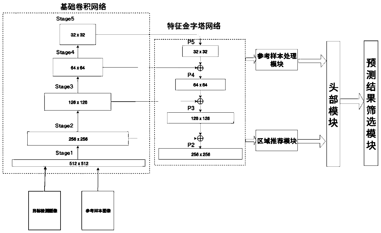
[0157] Such as Figure 2-9 As shown, the present embodiment provides a comparison detector, including a sequentially connected basic convolutional network, a feature pyramid network, a reference sample processing module, a region recommendation module, a head module and a prediction result screening module;
[0158] The basic convolutional network is constructed by a multi-layer convolutional module, which is specifically composed of several stages. As the stage increases, the resolution of the feature map decreases and the number of channels increases. Each stage consists of a certain number of convolutional layers. ;
[0159] The feature pyramid network is also constructed by a multi-layer convolution module, including the use of the basic convolutional network from the second stage to the last stage, and the last layer of features in each stage and the features above it. The corresponding feature m...
Embodiment 3
[0198] Example 3 The application example of the comparative detector described in Example 1 constructed by the method of Example 2 in the detection of cervical cancer cells
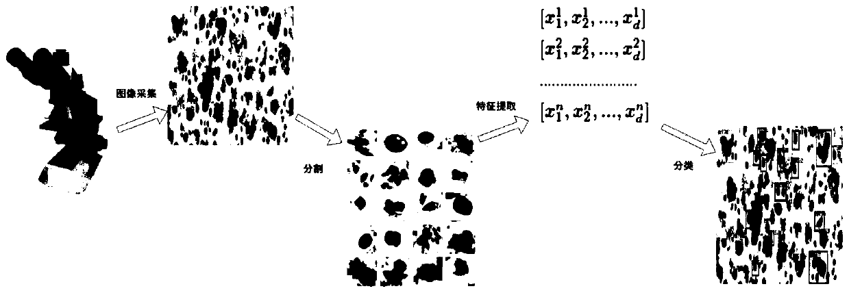
[0199] Such as Figure 2-9 As shown, the present embodiment provides a method for detecting cervical cancer cells based on the above-mentioned comparative detector, comprising the following steps:
[0200] Step 1. Construct the training set and test set of cervical microscopic images:
[0201] Collect microscopic images of the cervix and label the key components in the microscopic images of the cervix, and randomly select the microscopic images of the cervix after the labeling operation to construct a training set and a test set;
[0202] The operation of marking the key components in cervical microscopic images refers to marking the area where the key components in cervical microscopic images are located by systematically trained professionals (such as Figure 9 (a)), and record the center coordinate p...
PUM
 Abstract
Description
Claims
Application Information
 - R&D
- Intellectual Property
- Life Sciences
- Materials
- Tech Scout
- Unparalleled Data Quality
- Higher Quality Content
- 60% Fewer Hallucinations
Browse by: Latest US Patents, China's latest patents, Technical Efficacy Thesaurus, Application Domain, Technology Topic, Popular Technical Reports.
© 2025 PatSnap. All rights reserved.Legal|Privacy policy|Modern Slavery Act Transparency Statement|Sitemap|About US| Contact US: help@patsnap.com


