
Preparation method of separation tube for separating tumor cells

A technology of tumor cells and separation tubes, which is applied in the field of preparation of separation tubes, can solve the problems of inability to obtain tumor cells and unavoidable pollution of other cells, and achieve the effects of high purity, convenient operation, and avoiding pollution

Active Publication Date: 2019-07-19
JIANGSU COWIN BIOTECH CO LTD
View PDF7 Cites 2 Cited by
  • Summary
  • Abstract
  • Description
  • Claims
  • Application Information

AI Technical Summary

Problems solved by technology

However, directly using Percoll to separate circulating tumor cells or body cavity effusion tumor cells by density centrifugation method, while harvesting tumor cells, it will inevitably be polluted by other cells, and tumor cells with higher purity cannot be obtained.

Method used


Image

  • Preparation method of separation tube for separating tumor cells
  • Preparation method of separation tube for separating tumor cells
  • Preparation method of separation tube for separating tumor cells

Examples


Embodiment 1

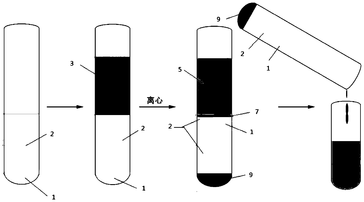
[0031] A method for preparing a separation tube for separating tumor cells, the method is used for separating tumor cells in body cavity effusions, such as figure 1 shown, including the following steps:

[0032] S1: First add 5mL of separation gel to a 50mL separation tube. At room temperature, set the centrifugal force to 1000g and centrifuge for 5 minutes to make the separation gel sink to the bottom of the separation tube. The separation gel has thixotropy characteristics and a density of 1.06-1.08 g / mL;

[0033] S2: Add 20mL of Percoll separation medium to the upper layer of the separation gel, the concentration of Percoll separation medium is 38-42%, and the corresponding density is 1.05-1.06g / ml;

[0034] S3: Slowly add 20 mL of body cavity fluid obtained by puncture of a tumor patient to the upper layer of Percoll separation medium, set the centrifugal force at 1000 g at room temperature, and centrifuge for 10 min, figure 1 As shown, non-target cells such as red blood...

Embodiment 2

[0039] A method for preparing a separation tube for isolating tumor cells, the method is used for isolating circulating tumor cells such as Figure 4 shown, including the following steps:

[0040] S1: First add 2mL of separation gel to a 15mL separation tube. At room temperature, set the centrifugal force to 1000g and centrifuge for 5 minutes to make the separation gel sink to the bottom of the separation tube. The separation gel has thixotropy characteristics and a density of 1.06-1.08 g / mL;

[0041] S2: Add 6mL of Percoll separation medium to the upper layer of the separation gel, the concentration of Percoll separation medium is 38-42%, and the corresponding density is 1.05-1.06g / ml;

[0042] S3: Slowly add 6mL of anticoagulated whole blood to the upper layer of Percoll separation medium, set the centrifugal force at 1000g at room temperature, and centrifuge for 10min, as Figure 4As shown, non-target cells such as red blood cells and white blood cells sink to the bottom ...


PUM


Abstract

The invention provides a preparation method of a separation tube for separating tumor cells, and belongs to the technical field of cell separation. The preparation method comprises the following stepsof firstly adding separation gel to the separation tube, performing centrifuging so as to enable the separation gel to sink to the bottom of the tube, then adding separation mediums, then slowly adding body cavity hydrops or anticoagulation whole blood to an upper layer of the separation mediums, performing centrifuging at room temperature, firstly sucking and discarding liquid at the topmost surface of the separation tube so as to remove cell debris, then dumping residual liquid on the separation gel layer into a collecting tube, then performing centrifuging at room temperature, performing cell re-suspending with PBS so as to obtain the tumor cells, wherein the density of the separation gel is 1.06-1.08g / mL, and the density of the separation mediums is 1.05-1.06g / mL. The preparation method disclosed by the invention can be used for separating and circulating the tumor cells from the tumor cells in the body cavity hydrops, the preparation method is simple, convenient and efficient, and the purity of the separated tumor cells is high.

Description

technical field [0001] The invention belongs to the technical field of cell separation, and in particular relates to a method for preparing a separation tube for separating tumor cells. Background technique [0002] Body cavity effusion refers to the effusion in the cavities around the organs of the human body, mainly including the pleural cavity, abdominal cavity, pelvic cavity and subarachnoid space. Body cavity effusions are associated with many types of malignancies, such as liver cancer, lung cancer, breast cancer, Koposi's sarcoma, and lymphoma, among others. Isolation and identification of tumor cells and their gene mutation types in body cavity fluid have important clinical significance for disease diagnosis, disease assessment and formulation of treatment strategies. The cell types in the body cavity fluid of tumor patients are relatively complex, mainly including red blood cells, white blood cells, and mesothelial cells, etc., and the number of tumor cells is rela...

Claims


Application Information

Patent Timeline
Patent Type & Authority Applications(China)
IPC IPC(8): C12N5/09
CPCC12N5/0693C12N2509/00
Inventor 王春香刘勇
Owner JIANGSU COWIN BIOTECH CO LTD
Learn More