
Method for preparing micron-sized single-particle multi-TEM slice sample in situ

An in-situ preparation, single-particle technology, applied in the fields of planetary science and planetary exploration, can solve the problems of large-sized micron-scale sample preparation, waste of sample resources, and difficulty in applying micron-scale powder samples, etc. The effect of overcoming area damage

Active Publication Date: 2019-07-26
INST OF GEOCHEM CHINESE ACADEMY OF SCI
View PDF8 Cites 6 Cited by
  • Summary
  • Abstract
  • Description
  • Claims
  • Application Information

AI Technical Summary

Problems solved by technology

[0006] In view of the waste of sample resources in the preparation of TEM thin slices by the existing conventional FIB technology, it is difficult to apply to micron-sized powder samples, and the single-particle sample preparation technology combined with the existing FIB and ultra-thin microtome cannot achieve in-situ extraction As well as the inability to prepare samples for larger micron-scale samples, the present invention provides a method for in-situ preparation of micron-scale single-particle multiple TEM flake samples, so as to achieve a sample size ranging from tens of microns to about 200 microns In-situ preparation of single-particle sample TEM thin slices, especially suitable for the preparation of TEM thin slices of rare extraterrestrial micron-scale powder samples

Method used


Image

  • Method for preparing micron-sized single-particle multi-TEM slice sample in situ
  • Method for preparing micron-sized single-particle multi-TEM slice sample in situ
  • Method for preparing micron-sized single-particle multi-TEM slice sample in situ

Examples


Embodiment 1

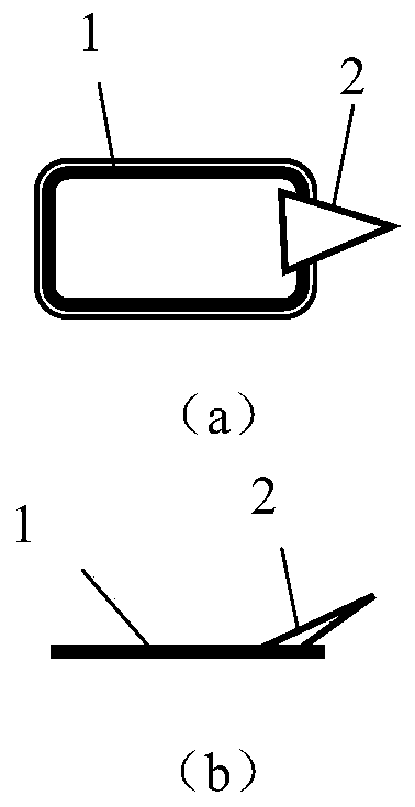
[0040] In this example, an optical microscope, a nanomanipulator and a double-beam electron microscope are combined to prepare micron-sized single-particle multiple TEM flake samples in situ, and the steps are as follows:

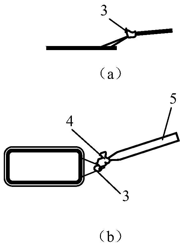
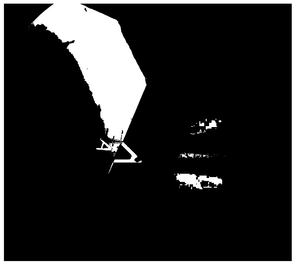
[0041] (1) if figure 2 As shown, manually add a little UV-curable glue on the needle tip, place it under an optical microscope, and use a nanomanipulator to place one end of an irregular single-particle sample with a maximum width of about 50 microns on the needle tip containing UV-curable glue, and the other end is suspended. , UV curable glue is cured by ultraviolet radiation to complete the partial suspension bonding and fixation of the single particle sample on the needle tip. The photo of using the nanomanipulator to bond the single particle sample to the needle tip containing UV-curable adhesive under the optical microscope is as follows: image 3 shown. The needle tip is an accessory of the atomic force microscope, which consists of a base and a t...

Embodiment 2

[0050] In this example, an optical microscope, a nanomanipulator and a double-beam electron microscope are combined to prepare micron-sized single-particle multiple TEM flake samples in situ, and the steps are as follows:

[0051] (1) Manually add a little UV-curable glue on the needle tip, place it under an optical microscope, and use a nanomanipulator to place one end of an irregular single-particle sample with a maximum width of about 40 microns on the needle tip containing UV-curable glue, and the other end is suspended , UV curable glue is cured by ultraviolet radiation to complete the partial suspension bonding and fixation of the single particle sample on the needle tip. The needle tip is an accessory of the atomic force microscope, which consists of a base and a triangular cantilevered needle tip located on the edge of the base. There is a certain angle between the cantilevered needle tip and the base, and the cantilevered needle tip has certain elasticity, such as fig...

Embodiment 3

[0065] In this example, an optical microscope, a nanomanipulator and a double-beam electron microscope are combined to prepare micron-sized single-particle multiple TEM flake samples in situ, and the steps are as follows:

[0066] (1) if figure 2 As shown, manually add a little UV-curable glue on the needle tip, place it under an optical microscope, use a nanomanipulator to place a part of the irregular single-particle sample with a maximum width of about 144 microns on the needle tip containing UV-curable glue, and the other part is suspended , UV curable glue is cured by ultraviolet radiation to complete the partial suspension bonding and fixation of the single particle sample on the needle tip. The needle tip is an accessory of the atomic force microscope, which consists of a base and a triangular cantilevered needle tip located on the edge of the base. There is a certain angle between the cantilevered needle tip and the base, and the cantilevered needle tip has certain el...


PUM

PropertyMeasurementUnit
Sizeaaaaaaaaaa
Widthaaaaaaaaaa
Thicknessaaaaaaaaaa

Abstract

The invention belongs to the field of planetary science and planetary detection, and provides a method for preparing a micron-sized single-particle multi-TEM slice sample in situ. The method comprisesthe following steps: adhering and fixing a single-particle sample on a needle point in a partially suspended mode, placing the single-particle sample on a sample table of a double-beam electron microscope, installing an FIB carrying net, sealing a sample cavity and vacuumizing; depositing a Pt layer on the surface of the single-particle sample; observing the single-particle sample from an ion beam interface of the double-beam electron microscope, selecting an interested region, performing denudation cutting processing on the single-particle sample by using FIB, cutting the interested region from the single-particle sample to obtain a slice, and bonding the slice with a nano manipulator configured by the double-beam electron microscope; adhering the slice on the nanometer manipulator to anFIB carrying net to enable the slice to be vertical to the sample table, cutting off the connection between the slice and the nanometer manipulator, and thinning the slice by using the FIB to preparea TEM slice sample; and repeating the cutting and thinning operations to prepare single particle samples into a plurality of TEM sheet samples.

Description

technical field [0001] The invention belongs to the fields of planetary science and planetary exploration, and relates to a method for preparing TEM flakes by using a double-beam electron microscope, and more specifically, relates to a method for preparing micron-sized single-particle multiple TEM flake samples in situ. Background technique [0002] In the past ten years, the nanoprocessing technology of focused ion beam-electron beam (FIB / SEM) electron microscope (dual-beam electron microscope), as an important tool for transmission electron microscope (TEM) sample preparation, has been widely used in materials, biology, and geochemistry. , planetary science, and geology. Compared with other processes, such as ultramicrotome, ion polisher, etc., the biggest advantage of dual-beam electron microscopy is that it can prepare TEM thin-section samples in situ. The technique has received widespread attention from a growing number of researchers in the fields of planetary science...

Claims


Application Information

Patent Timeline
IPC IPC(8): G01N23/2202
CPCG01N23/2202G01N1/286G01N1/32G01N2001/045H01J2237/208H01J2237/2583H01J2237/31745H01J2237/31749
Inventor 李瑞李阳金宏李雄耀王世杰
Owner INST OF GEOCHEM CHINESE ACADEMY OF SCI
Learn More