SPECT and PET image correction method and device and electronic equipment
An image correction and image technology, which is applied in the field of image processing, can solve the problems of low accuracy of SPECT and PET images, and achieve the effect of solving low accuracy, improving accuracy, and effectively correcting processing
- Summary
- Abstract
- Description
- Claims
- Application Information
AI Technical Summary
Problems solved by technology
Method used
Image
Examples
Embodiment 1
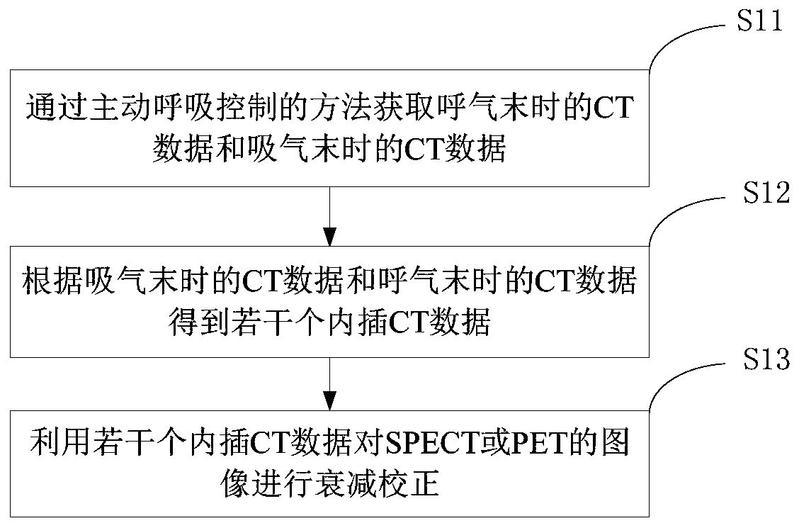
[0064] An image correction method for SPECT and PET provided in the embodiment of the present application, such as figure 1 As shown, the method includes:
[0065] S11: Obtain the CT data at the end of expiration and the CT data at the end of inspiration by means of active breathing control.
[0066] The method of respiratory gating refers to the imaging technology that utilizes the thoracic movement law during respiratory movement to eliminate the impact of respiratory movement on the image quality of ultrasound, X-ray, CT, and magnetic resonance imaging. It can also be called respiratory gating. imaging technology. Specifically, during the imaging process, imaging equipment (such as ultrasound, X-ray, CT, magnetic resonance imaging, etc.) is prone to produce artifacts due to the movement of tissues or organs (such as breathing, heartbeat, etc.), which will reduce the resolution of the image. rate and diagnostic value.
[0067] In this embodiment, the average attenuation m...
Embodiment 2
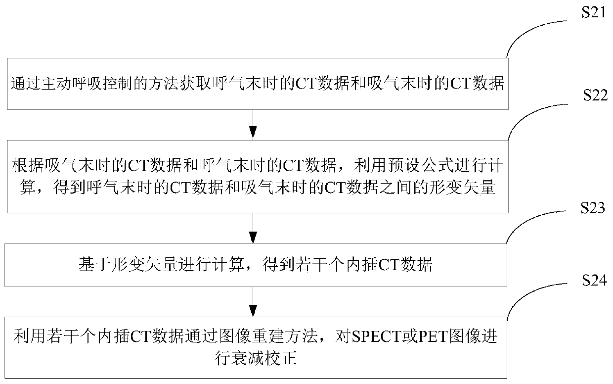
[0078] An image correction method for SPECT and PET provided in the embodiment of the present application, such as figure 2 shown, including:
[0079] S21: Obtain the CT data at the end of expiration and the CT data at the end of inspiration by means of active breathing control.
[0080] In this step, the CT data at the end of expiration and the CT data at the end of inspiration are obtained from the CT images. Wherein, the CT image may be a CT image of lungs, heart, liver and other parts. This embodiment is described by taking the SPECT image, the PET image and the CT image as heart images as an example.
[0081] S22: According to the CT data at the end of inspiration and the CT data at the end of expiration, calculate using a preset formula to obtain the deformation vector between the CT data at the end of expiration and the CT data at the end of inspiration, wherein, The default formula is:
[0082] Among them, CF is the cost function, F iis the CT data at the end o...
Embodiment 3
[0133] An image correction device for SPECT and PET provided in the embodiment of the present application, such as Figure 16 As shown, the image correction device 3 for SPECT and PET includes: a first acquisition module 31 , a second acquisition module 32 and a correction module 33 .
[0134] The first acquiring module 31 is used to acquire the CT data at the end of expiration and the CT data at the end of inspiration by means of active breathing control. The second acquisition module 32 is used to obtain several interpolated CT data according to the CT data at the end of inspiration and the CT data at the end of expiration. The correction module 33 is used to perform attenuation correction on respiratory-gated SPECT and PET images using several interpolated CT data.
[0135] The second acquisition module is specifically used for: firstly, according to the CT data at the end of inspiration and the CT data at the end of expiration, use a preset formula to calculate, and obtai...
PUM
 Abstract
Description
Claims
Application Information
 - R&D
- Intellectual Property
- Life Sciences
- Materials
- Tech Scout
- Unparalleled Data Quality
- Higher Quality Content
- 60% Fewer Hallucinations
Browse by: Latest US Patents, China's latest patents, Technical Efficacy Thesaurus, Application Domain, Technology Topic, Popular Technical Reports.
© 2025 PatSnap. All rights reserved.Legal|Privacy policy|Modern Slavery Act Transparency Statement|Sitemap|About US| Contact US: help@patsnap.com


