
Method for inducing amniotic fluid mesenchymal stem cells to differentiate into cardiomyocytes in vitro

A technique for stem cells and cardiomyocytes, applied in the field of stem cells, can solve the problems of inability to repair myocardial tissue in a timely and effective manner, afterload mismatch, and long time-consuming repair.

Inactive Publication Date: 2019-08-16
南京乐扬医药科技有限公司
View PDF0 Cites 0 Cited by
  • Summary
  • Abstract
  • Description
  • Claims
  • Application Information

AI Technical Summary

Problems solved by technology

After myocardial necrosis occurs in heart disease, although the myocardial progenitor cells contained in the myocardium will undergo division and hyperplasia for some compensatory repair, the number is small and the proliferation ability is low, and the repair takes a long time. The demand for regeneration of myocardial tissue cannot be met in the short term, and the necrotic myocardium can only be replaced by fibrous scar tissue in the end, resulting in ventricular remodeling, changes in ventricular structure, resulting in afterload mismatch and left ventricular dysfunction, resulting in a difficult-to-treat end-stage congestive heart failure

Method used


Image

  • Method for inducing amniotic fluid mesenchymal stem cells to differentiate into cardiomyocytes in vitro
  • Method for inducing amniotic fluid mesenchymal stem cells to differentiate into cardiomyocytes in vitro
  • Method for inducing amniotic fluid mesenchymal stem cells to differentiate into cardiomyocytes in vitro

Examples

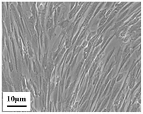

Embodiment Construction

[0022] 1. Experimental method

[0023] 1. Culture of human amniotic fluid mesenchymal stem cells

[0024] Human amniotic fluid mesenchymal stem cells (Shanghai Yaji Biotech) were resuscitated with low-sugar DMEM medium (U.S. Gibco) containing 20% ​​calf serum (Shanghai Suer Biology) and 2ng / mL basic fibroblast growth factor (U.S. Sigma) At volume fraction 5% CO 2 , Cultivated in a 37°C incubator, and the cells were passaged at a ratio of 1:4 when the cells reached 80% confluence.

[0025] 2. Identification of human amniotic fluid mesenchymal stem cells

[0026] 2.1 Morphological observation

[0027] Human amniotic fluid mesenchymal stem cells in the logarithmic growth phase were placed under an inverted microscope to observe the cell morphology.

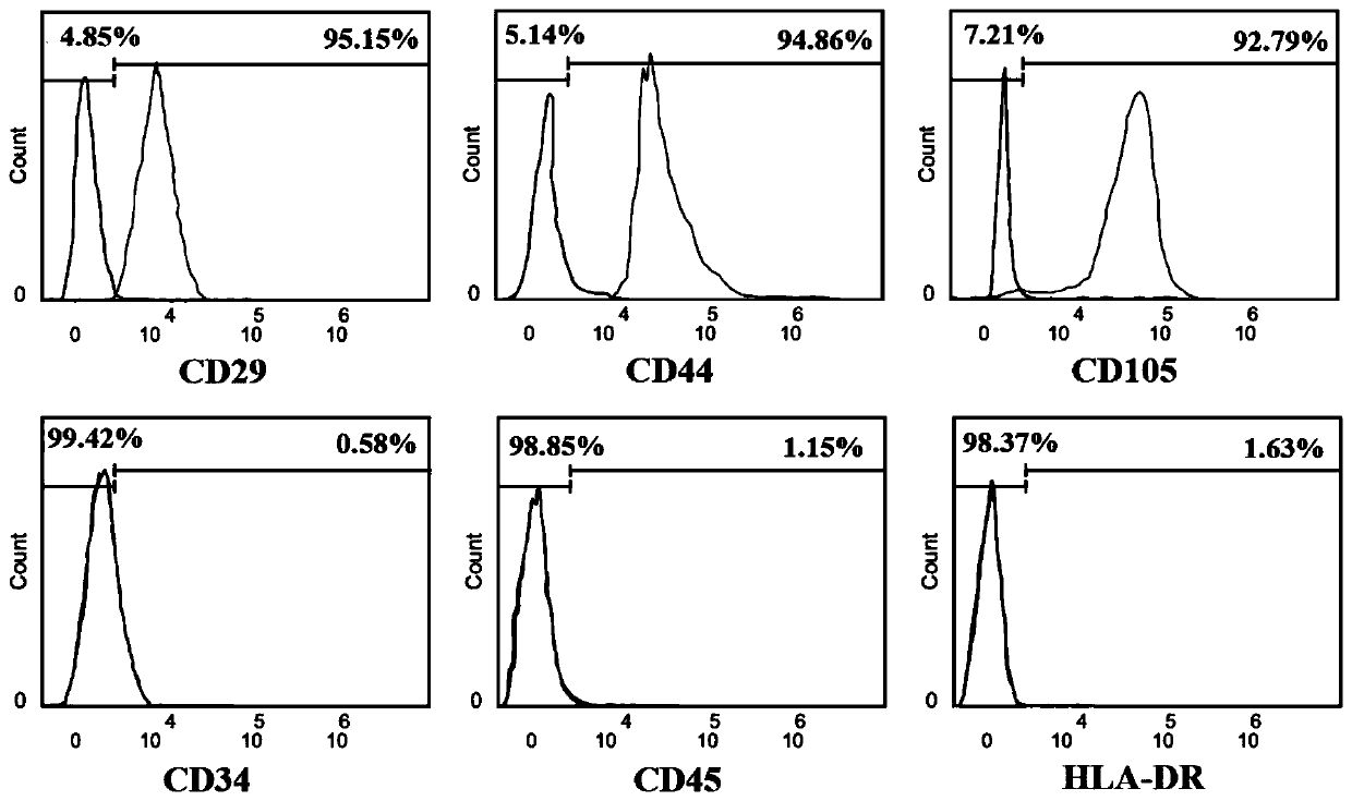
[0028] 2.2 Detection of cell surface markers

[0029] Human amniotic fluid mesenchymal stem cells in the logarithmic growth phase were taken, washed 3 times with PBS, and prepared with PBS at a concentration of 2×10 6 / mL of ce...


PUM


Abstract

The invention discloses a method for inducing amniotic fluid mesenchymal stem cells to differentiate into cardiomyocytes in vitro. CTnI is a typical protein in ventricular and atrial cells and is an important characteristic protein of cardiomyocytes; GATA4 is an important transcriptional regulatory factor for cardiac gene expression, jointly regulates the normal development of a heart, the expression of functional genes and the pathological process of cardiac hypertrophy through interaction with other transcriptional regulatory factors, and is also an important characteristic protein of the cardiomyocytes. Through mRNA and protein expression levels of the cTnI and GATA4, the human amniotic fluid mesenchymal stem cells can be observed to be obviously differentiated into the cardiomyocytes under induction of propylgallate. Therefore, propylgallate can be used for inducing the human amniotic fluid mesenchymal stem cells to differentiate into the cardiomyocytes in vitro. The method provided by the invention can effectively induce the human amniotic fluid mesenchymal stem cells to differentiate into the cardiomyocytes.

Description

technical field [0001] The invention belongs to the field of stem cells, and relates to the induced differentiation of stem cells, in particular to a method for inducing the differentiation of amniotic fluid mesenchymal stem cells into cardiomyocytes in vitro. Background technique [0002] Stem cells are a class of cells with self-renewal and multilineage differentiation capabilities. It is because of its regenerative potential and plasticity that it is widely used in regenerative medicine and stem cell research. Stem cells hold promise for many diseases that currently lack effective treatments, including neurological and cardiovascular diseases. [0003] Mesenchymal stem cells are multipotent stromal cells isolated from adult and fetal tissues, defined as adherent fibroblast-like cells. Mesenchymal stem cells also have an intrinsic homing ability, can migrate to damaged tissues, and actively participate in tissue repair. In addition, mesenchymal stem cells have strong im...

Claims


Application Information

Patent Timeline
IPC IPC(8): C12N5/077C12N5/0775
CPCC12N5/0657C12N2501/115C12N2501/999C12N2506/1392
Inventor 马春华陆扬刘燕毕晓娟
Owner 南京乐扬医药科技有限公司
Learn More