Blocking type PD-L1 camel-source single-domain antibody and use thereof

A PD-L1, single-domain antibody technology, applied in the field of biomedicine or biopharmaceuticals, can solve problems such as the reduction of T cell immune effect

Active Publication Date: 2019-08-20
SHANGHAI NOVAMAB BIOPHARM CO LTD
View PDF6 Cites 9 Cited by
  • Summary
  • Abstract
  • Description
  • Claims
  • Application Information

AI Technical Summary

Problems solved by technology

However, activation of the PD-1 / PD-L1 signaling pathway changes the local microenvironment of the tumor and reduces the immune effect of T cells, thereby mediating tumor immune escape and promoting tumor growth

Method used


Image

  • Blocking type PD-L1 camel-source single-domain antibody and use thereof
  • Blocking type PD-L1 camel-source single-domain antibody and use thereof
  • Blocking type PD-L1 camel-source single-domain antibody and use thereof

Examples


Embodiment 1

[0139] Example 1: Construction, screening and identification of PD-L1 single domain antibody library

[0140] Library construction:

[0141] (1) Mix 1 mg of hPD-L1(ECD)-Fc antigen with Freund's adjuvant in equal volume, immunize a Xinjiang Bactrian camel once a week, and immunize 7 times in total to stimulate B cells to express antigen-specific single domain Antibody; (2) After 7 times of immunization, extract 100mL camel peripheral blood lymphocytes and extract total RNA; (3) Synthesize cDNA and amplify VHH by nested PCR; (4) Use restriction enzymes PstI and NotI Cut 20 μg of pMECS phage display vector and 10 μg of VHH and ligate the two fragments; (5) Transform the ligation product into electroporation-competent cells TG1 to construct a PD-L1 single domain antibody library.

[0142] Antibody Screening:

[0143] (1) 10 μg ghPD-L1(ECD)-Fc antigen (10 μg Fc inNaHCO 3 As a control) coupled to NUNC microtiter plates, placed overnight at 4°C; (2) the next day, 100 μL of 0.1% BS...

Embodiment 2

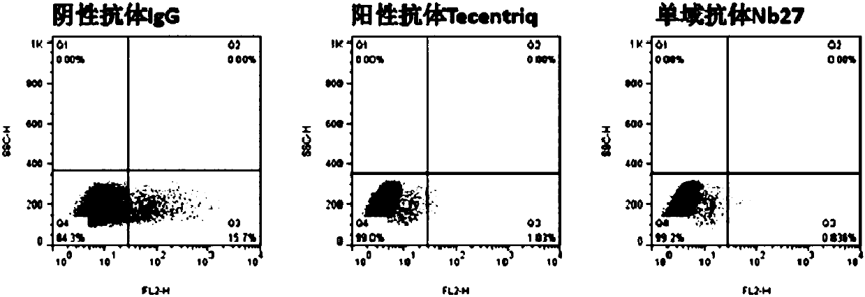
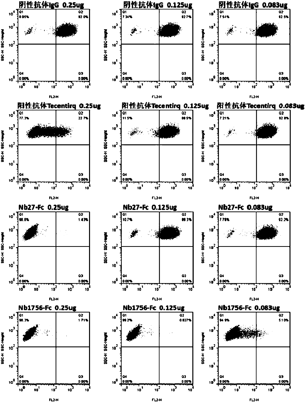
[0146] Example 2: Preliminary identification of blocking function of single domain antibody by flow cytometry

[0147] (1) Preparation of hPD-1(ECD)-Biotin protein (the preparation method of hPD-1(ECD) is the same as in Example 1), and the method of protein biotinylation refers to the instructions of biotin reagent; (2) Transient PD-L1 gene Express PD-L1 protein on the cell surface of HEK293F cells; (3) Prepare crude lysate of PD-L1 single domain antibody TG1 strain, the preparation method is the same as that of Zhu Min et al., Nanoscale Res Lett., 2014Sep26; 9(1) : 528; (4) Each sample takes 1×10 6 HEK293F cells transiently transfected with PD-L1 were resuspended in 0.5% BSA-PB Sbuffer, 100 μL of the above crude extract was added, and negative control (hIgG1) and positive control (Tecentriq) were set at the same time, and 5 μghPD-1 (ECD) was added to each well -Fc-Biotin, incubated at 4°C for 20 min; (5) Wash the cells twice with PBS, add eBioscience SA-PE, incubate at 4°C f...

Embodiment 3

[0148] Example 3: Humanized transformation of PD-L1 blocking single domain antibody

[0149] (1) First use the PD-L1 single domain antibody sequence shown in SEQ ID NO.:8 as a template to search for homologous structures in the structural database, and take the structure where Evalue=0.0 and sequence identity ≥ 70%; (2 ) Structural comparison of these structures, and according to the crystal structure resolution size and the constructed phylogenetic tree, multi-template homology modeling based on the PD-L1 single domain antibody sequence shown in SEQ ID NO.:8, and then According to the ranking of the scoring function, select the structure with the lowest molpdf; (3) For the optimal structure modeled, use the ProtSA server to calculate the solvent accessibility of the residue, that is, the solvent accessibility of the folded state of the residue relative to the unfolded state. The ratio of the contact area is the criterion, and the residues greater than 40% are taken as the res...


PUM


Abstract

The invention relates to a camel-source single-domain antibody. The antibody specifically recognizes programmed death receptor-ligand 1(PD-L1), the interaction of PD-L1 and the receptor PD-1 thereof can be effectively blocked, and the blocking effect is better than that of atezolizumab which is sold in the market. Specifically speaking, the invention discloses a PD-L1 camel-source single-domain antibody, a coding sequence of derived protein of the PD-L1 camel-source single-domain antibody, and a preparation method and use of the derived protein.

Description

technical field [0001] The present invention relates to the technical field of biomedicine or biopharmaceuticals, and more specifically relates to an anti-PD-L1 camel-derived single domain antibody and its application. Background technique [0002] Tumor immune escape means that tumor cells evade collective immune recognition and attack through changes in themselves or the microenvironment. Due to its advantages of less damage and effective control of tumor growth and metastasis, tumor immunotherapy has become an urgent need for clinical tumor treatment. Programmed death 1 / programmed death 1 ligand 1 (PD-1 / PD-L1), as a pair of negative immune co-stimulatory molecules, participates in tumor immune evasion. PD-1 / PD-L1 is a new member of the CD28 / B7 co-stimulatory molecule superfamily, which can mediate negative co-stimulatory signals, effectively inhibit the function and proliferation of T and B cells, and reduce the cytokines IL-2, IL- 10 and IFN-γ secretion, the immune reg...

Claims


Application Information

Patent Timeline
IPC IPC(8): C07K16/28C07K16/46C07K19/00
CPCC07K16/2827C07K16/46C07K2317/569C07K2319/30A61P35/00C12N15/1037A61K2039/505C07K2317/22C07K2317/24C07K2317/92C07K2317/94C07K2317/35C07K2317/33C07K2317/34C07K2317/76C12Q2565/626C07K2317/565C07K2317/567
Inventor 万亚坤朱敏沈晓宁
Owner SHANGHAI NOVAMAB BIOPHARM CO LTD
Why Eureka
Try Eureka