Primary isolation and culture method of human umbilical vein endothelium

A technology for separating and culturing umbilical veins, applied in the biological field, can solve the problems of various reagents, decreased cell viability, aging, shrinking and death, etc., to avoid vascular smooth muscle shedding, improve feasibility, and good cell viability.

Inactive Publication Date: 2019-09-03
XI AN JIAOTONG UNIV
View PDF3 Cites 2 Cited by
  • Summary
  • Abstract
  • Description
  • Claims
  • Application Information

AI Technical Summary

Problems solved by technology

At present, there are commercial disposable primary endothelial cells on the market, but the companies are mixed, the real source of cells is lack of supervision, the market is mixed, and its price is expensive. Therefore, it is a convenient, efficient, economical and stable way to extract and cultivate primary umbilical The method of venous endothelium has important scientific research value and economic significance
[0003] The existing primary isolation and culture methods of umbilical vein endothelium have extremely high requirements on the timeliness of the specimens. If the umbilical cord is isolated for more than one hour and the separation has not started, the cell viability will be severely reduced, which will greatly affect the cell viability and passage times after inoculation. Therefore, the decline in the viability of the specimens caused by the transfer of freshly isolated specimens from the delivery room to the laboratory is the first difficulty in the separation; and the types of reagents required are complicated and expensive, and the extracted primary cells are unstable. The number of times is often less than 3 generations, aging, shrinkage and death, which is difficult to meet the requirements of basic scientific research
[0004] Existing schemes have extremely high requirements on the timeliness of specimens. After the umbilical cord is isolated, it is placed in normal saline for storage. If the separation has not started after more than one hour, the cell viability will seriously decline, which greatly affects the cell viability and passage after inoculation. The number of times the umbilical cord is transferred from the delivery room to the laboratory will lead to a decrease in cell viability. Therefore, it is urgent to improve the formula of the umbilical cord storage solution to extend the storage time of the specimen as much as possible while ensuring cell viability.
Even if the specimen can be separated quickly after isolation, the special medium set for endothelial cells is expensive, the reagents required for the culture process are various, and the extracted primary cells are unstable, and the number of cell passages is usually less than 3 generations. Aging, shrinking and death, it is difficult to meet the requirements of basic scientific research

Method used


Image

  • Primary isolation and culture method of human umbilical vein endothelium
  • Primary isolation and culture method of human umbilical vein endothelium
  • Primary isolation and culture method of human umbilical vein endothelium

Examples


Embodiment 1

[0062] ①Umbilical cords of healthy full-term fetuses were collected. Before the umbilical cord is collected, it is necessary to review the results of the maternal examination to exclude the maternal with infectious diseases (AIDS, hepatitis B, hepatitis C, syphilis, etc.) to ensure the safety of the experimental operators;

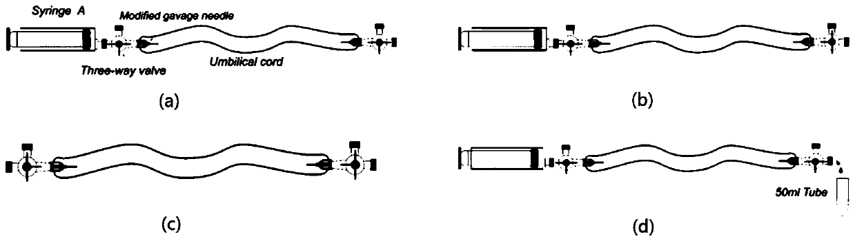
[0063] ②Take a 15cm-long umbilical cord in a sterile environment, cut off the hemostatic clamp marks at both ends of the umbilical cord, gently squeeze out the residual blood in the blood vessel, put the umbilical cord into the storage solution, place it on ice and transport it back to the laboratory;

[0064] ③Add 3mL 0.1% gelatin solution to the T25 culture flask, put it in 36.5℃, CO 2 4.5% concentration in the incubator for 30 minutes, pour off the gelatin solution before inoculation, and rinse the wall of the culture bottle with 2mL sterile PBS solution;

[0065] ④Transfer the umbilical cord to the sterilized waist plate in the ultra-clean bench, wipe...

Embodiment 2

[0071] ①Collect the umbilical cord of a healthy full-term fetus; before the collection of the umbilical cord, it is necessary to review the results of the maternal examination and exclude the maternal with infectious diseases (AIDS, hepatitis B, hepatitis C, syphilis, etc.) to ensure the safety of the experimental operators;

[0072] ②Take a 20cm-long umbilical cord in a sterile environment, cut off the hemostatic clamp marks at both ends of the umbilical cord, gently squeeze out the residual blood in the blood vessel, put the umbilical cord into the storage solution, place it on ice and transport it back to the laboratory;

[0073] ③Add 3mL 0.1% gelatin solution to the T25 culture flask, put it in 37.5℃, CO 2The concentration is 5.5% in the incubator for 30 minutes, pour off the gelatin solution before inoculation, and rinse the wall of the culture bottle with 2mL PBS solution;

[0074] ④Transfer the umbilical cord to the sterilized waist plate in the ultra-clean bench, wipe ...

Embodiment 3

[0080] ①Collect the umbilical cord of a healthy full-term fetus; before the collection of the umbilical cord, it is necessary to review the results of the maternal examination and exclude the maternal with infectious diseases (AIDS, hepatitis B, hepatitis C, syphilis, etc.) to ensure the safety of the experimental operators;

[0081] ②Take a umbilical cord with a length of 30 cm under a sterile environment, cut off the hemostatic clamp marks at both ends of the umbilical cord, gently squeeze out the residual blood in the blood vessel, put the umbilical cord into the storage solution, place it on ice and transport it back to the laboratory;

[0082] ③ Add 3mL 0.1% gelatin solution to the T25 culture flask, put it in 37℃, CO 2 The concentration is 5% in the incubator for 30 minutes, pour off the gelatin solution before inoculation, and rinse the wall of the culture bottle with 2mL PBS solution;

[0083] ④Transfer the umbilical cord to the sterilized waist plate in the ultra-clea...


PUM

PropertyMeasurementUnit
volumeaaaaaaaaaa

Abstract

The invention discloses a primary isolation and culture method for human umbilical vein endothelium, after collecting postoperative umbilical cord, collagenase is added to separate and extract umbilical vein endothelial cells to prepare a single cell suspension, the P0 generation cells are cultured, and the P0 generation cells are cultured to the P2 generation cells by using a complete medium based on the M199 medium; P2 cells are digested with trypsin, inoculated into a T75 flask, and cultured in a complete medium based on the M199 medium, and when the cell fusion degree is 80 to 90%, P3 cells of human umbilical vein endothelial cells after expansion and culture are obtained. The method can ensure the cell viability while extending the umbilical cord storage time to 6 hours.

Description

technical field [0001] The invention belongs to the field of biotechnology, and in particular relates to a primary isolation and culture method of human umbilical vein endothelium. Background technique [0002] Endothelial cells are a single layer of flat, phosphorous epithelial cells that line the inner surface of blood vessels, between the blood and the vessel wall or tissue. In the past, scholars generally believed that endothelial cells constituted a simple mechanical barrier. However, with the development of science, people's understanding of endothelial cells continued to deepen, and endothelial cells played an important role in maintaining the homeostasis of the body. In the body, endothelial cells can release a variety of active mediators, regulate vascular tension, mediate inflammatory reactions, participate in various pathophysiological processes such as angiogenesis and thrombus formation. Endothelial cells have become an important research tool in the field of l...

Claims


Application Information

Patent Timeline
Patent Type & Authority Applications(China)
IPC IPC(8): C12N5/071
CPCC12N5/069C12N2500/12C12N2500/14C12N2500/16C12N2500/24C12N2500/32C12N2500/35C12N2500/36C12N2500/38C12N2500/40C12N2509/00
Inventor 刘进军王政孙雨瑶赵弓枭李雨蓓
Owner XI AN JIAOTONG UNIV
Why Eureka
Try Eureka