Precise lung cancer tissue location method based on SVM model and combining image

A precise positioning and image pairing technology, applied in the field of computer vision and pattern recognition, to achieve the effect of simple calculation, high precision and easy to use

Inactive Publication Date: 2019-09-06
TIANJIN UNIVERSITY OF TECHNOLOGY
View PDF3 Cites 3 Cited by
  • Summary
  • Abstract
  • Description
  • Claims
  • Application Information

AI Technical Summary

Problems solved by technology

Raman spectroscopy has strong specificity, reflecting the changes in the biochem...

Method used


Image

  • Precise lung cancer tissue location method based on SVM model and combining image
  • Precise lung cancer tissue location method based on SVM model and combining image
  • Precise lung cancer tissue location method based on SVM model and combining image

Examples


Embodiment Construction

[0036] As shown in the figure: a method for precise positioning of lung cancer tissue based on SVM model combined with images, including the following steps:

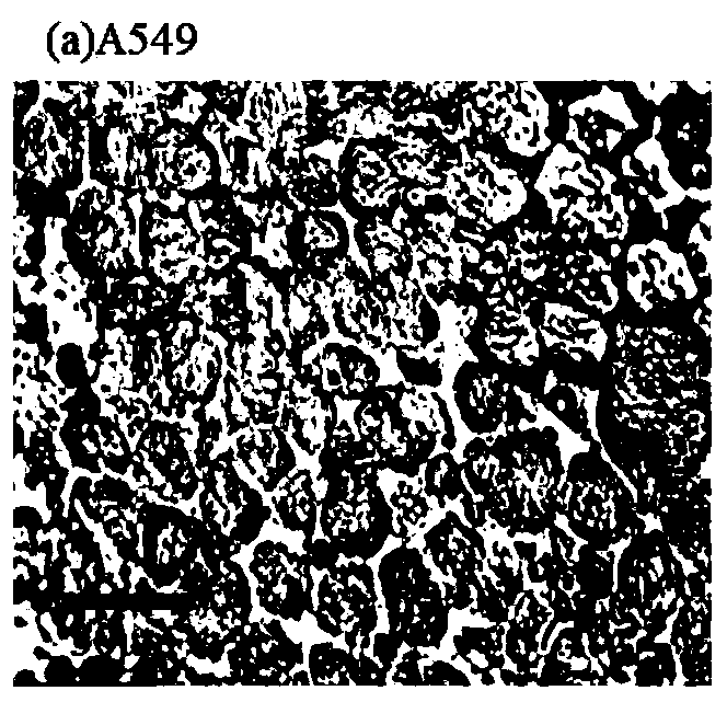
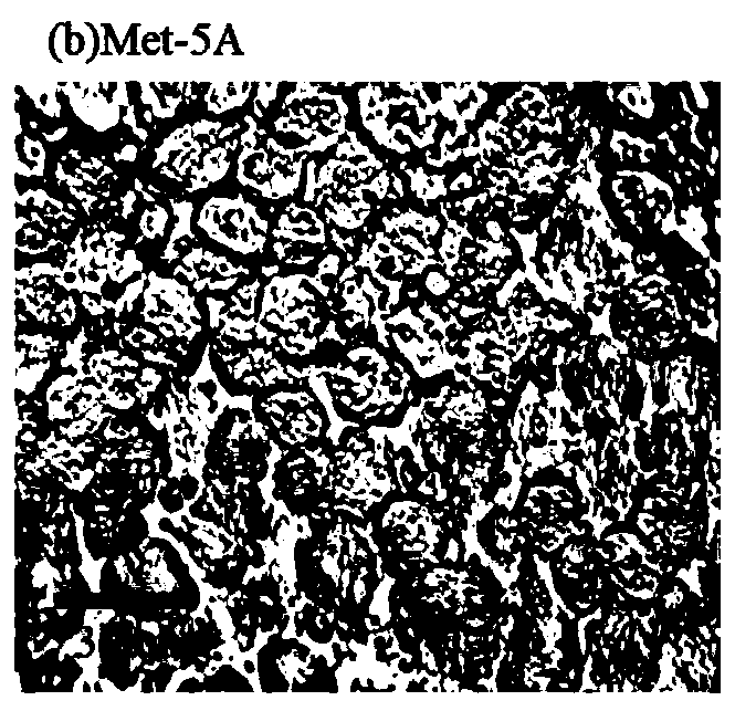
[0037] ①Cultivate two kinds of cells: lung adenocarcinoma cell line A549, pleural mesothelial cell line Met-5A, and the culture form is cell mass. The culture conditions are that the lung adenocarcinoma cell line A549 uses DMEM basal medium (containing 10% fetal bovine serum, 1% penicillin-streptomycin double antibody), and the pleural mesothelial cell line Met-5A uses DMEM high-glucose medium DMEM-H (containing 10% fetal bovine serum) at 37°C, 5% CO 2 Cultivate in an incubator, collect into a 15ml sterile centrifuge tube after cultivation, after washing twice with phosphate buffered saline (PBS), centrifuge to pellet the cells, centrifuge at 4500rpm for 10min, discard all the supernatant, and collect the cells for convenience observe.

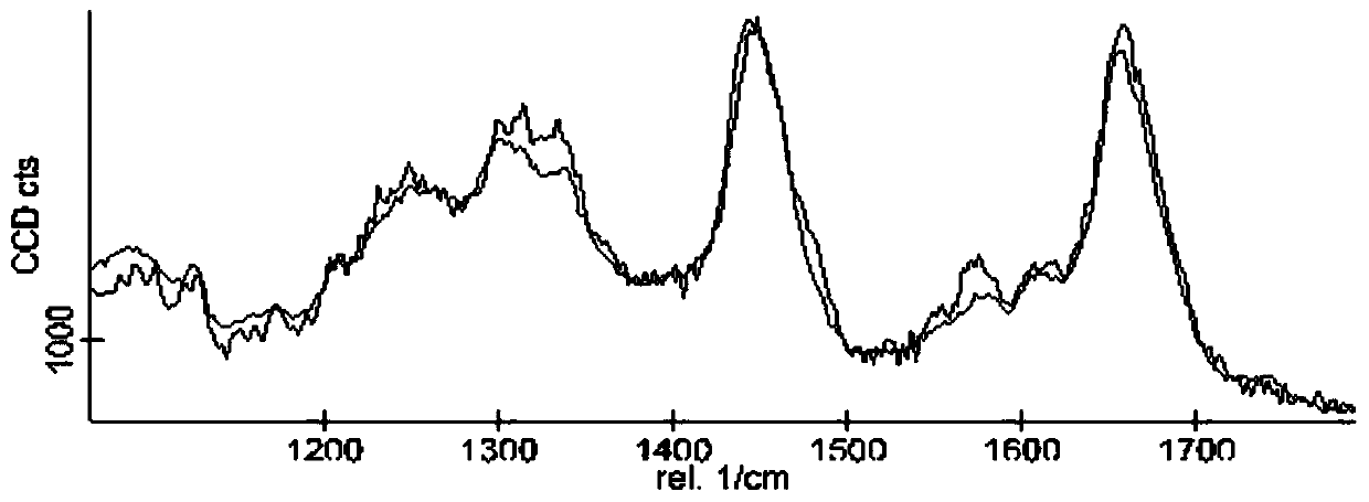
[0038] ②Put the cultured cell mass on a glass slide, and measure the spectrum of ...


PUM


Abstract

The invention relates to a precise lung cancer tissue location method which is based on an SVM model and combines with an image. The method comprises the following steps: (1) culturing two kinds of cell lines; (2) placing the two cultured cell lines on slides, and measuring Raman spectra of cells by adopting a confocal Raman spectrometer; (3) preprocessing the measured Raman spectra of the cells;(4) performing characteristic extraction on the preprocessed Raman spectra of the cells, wherein the extracted characteristics are characteristic peak positions and intensity ratios of the characteristic peak positions; (5) applying correlation analysis among multiple variables to the extracted characteristics; (6) for the characteristics extracted in the steps (4) and (5), performing classification recognition on spectral data by combining an SVM classifier; and (7) selecting a residual sample and testing, thus obtaining precision, sensitivity and specificity of the cells, and for a sample classified mistakenly, further distinguishing by utilizing a dyed image or Raman imaging. The method provided by the invention can eliminate the phenomenon that recognition rate is relatively low due toan error in an experiment or a sample culturing process.

Description

technical field [0001] The invention belongs to the field of computer vision and pattern recognition, in particular to a precise positioning method for lung cancer tissue based on an SVM model combined with images. Background technique [0002] Recognizing and locating objects is an important research content in the field of computer vision and pattern recognition. As a branch of object detection, the classification of cancer cells is a special case of object detection. Cells are a special kind of matter, which not only has universality but also has various particularities. Therefore, biometric detection has broad scientific research value and application prospects, and has very important research significance in medicine. [0003] Currently, fluorescent labeling is mainly used to identify cell types due to its specificity. Fluorescent labeling is based on the specific combination of antigens and antibodies. This method can easily damage the original physiological activity...

Claims


Application Information

Patent Timeline
IPC IPC(8): G01N21/65G06K9/00G06K9/62
CPCG01N21/65G06V20/698G06F18/2411
Inventor 赵萌闫静石凡陈胜勇
Owner TIANJIN UNIVERSITY OF TECHNOLOGY
Why Eureka
Try Eureka