Endoscope and endoscope system

An endoscope and imaging system technology, applied in the field of endoscope, can solve the problems that optical endoscope cannot obtain deep tissue lesion information, limit the completeness and accuracy of diagnosis, human digestive tract collision and friction, etc., and achieve reduction Check the blind area, the tolerance is convenient, and the effect of reducing the missed detection rate

Inactive Publication Date: 2019-10-25
SHENZHEN INST OF ADVANCED TECH
View PDF18 Cites 10 Cited by
  • Summary
  • Abstract
  • Description
  • Claims
  • Application Information

AI Technical Summary

Problems solved by technology

[0004] In view of this, the embodiment of the present invention provides an endoscope and an endoscope system to solve the problem that the existing optical endoscope cannot obtain the lesion information in the deep tissue, which limits the completeness and accuracy of the diagnosis, and is easy to It will cause collision and friction to the human digestive tract, which will bring discomfort to the patient

Method used


Image

  • Endoscope and endoscope system
  • Endoscope and endoscope system
  • Endoscope and endoscope system

Examples


Embodiment 1

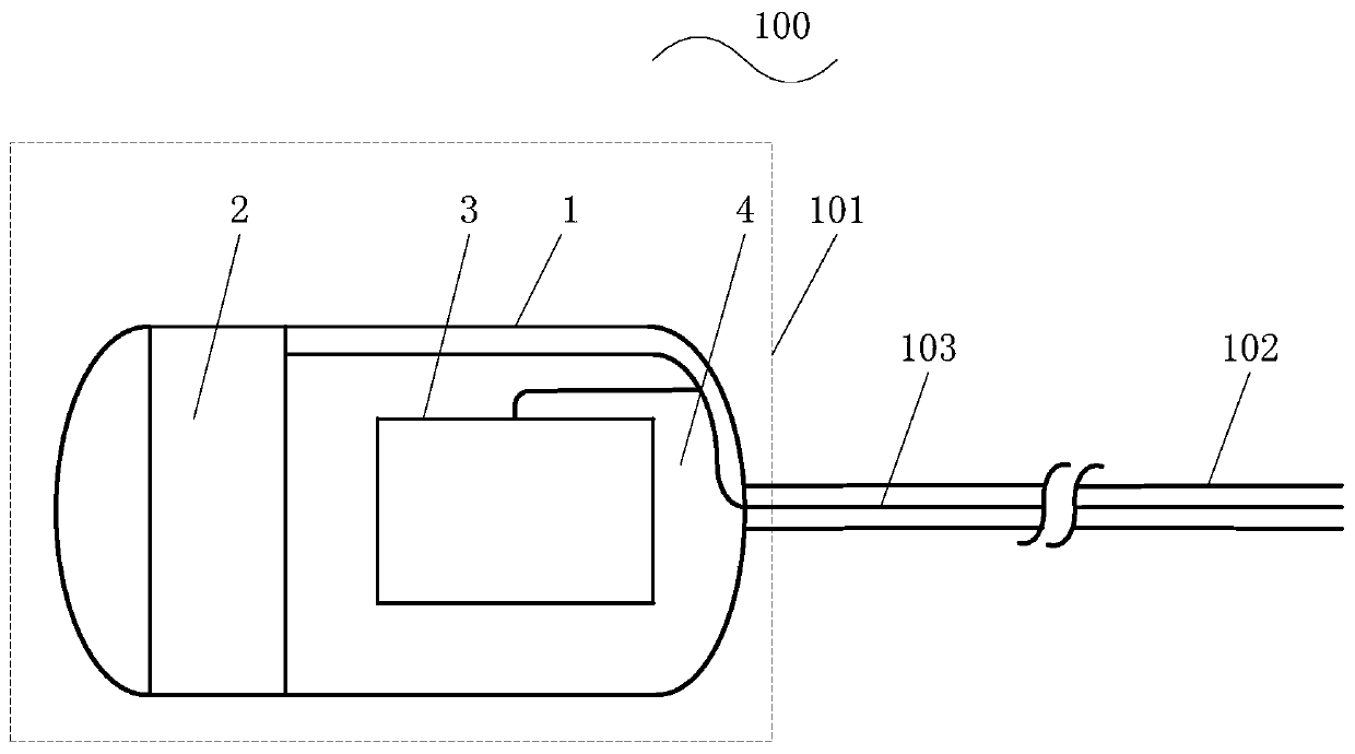
[0054] Such as figure 1 As shown, this embodiment provides an endoscope 100, including an endoscope main body 101, a flexible catheter 102 and a signal line 103, and the endoscope main body 101 includes a housing 1, a camera module 2 and an ultrasonic imaging module 3;

[0055] The endoscope body 101 is capsule-shaped and the outer diameter of the endoscope body 101 is larger than the outer diameter of the flexible catheter 102;

[0056] The camera module 2 and the ultrasonic imaging module 3 are arranged inside the casing 1, the flexible conduit 102 is sleeved outside the signal line 103 and connected to the casing 1, and one end of the signal line 103 is connected to the camera module 2 and the ultrasonic imaging module 3 , and the other end is used to connect with the imaging system.

[0057] In application, the main body of the endoscope can also be set in any other shape that is easy to swallow according to actual needs, for example, a spherical shape, an ellipsoid shape...

Embodiment 2

[0079] Such as Figure 4 As shown, in this embodiment, the endoscope 100 further includes a magnetic positioning component 4 , and the magnetic positioning component 4 is disposed on the casing 1 .

[0080] In application, the magnetic positioning part can be arranged inside or outside the housing, and the shape and size of the magnetic positioning part can be set according to actual needs. semi-elliptical.

[0081] Such as Figure 4 As shown, the magnetic positioning component 4 is exemplarily shown to be disposed on the outside of the housing 1 near the end of the flexible conduit 102 , and is semi-elliptical in shape and size with the same shape and size as the end of the casing near the flexible conduit.

[0082] In this embodiment, the magnetic positioning component 4 is used to drive the endoscope main body 101 to move into the human digestive tract under the magnetic attraction of the magnetic attraction component when the flexible catheter 102 and the signal line 103...

Embodiment 3

[0087] Such as Image 6 As shown, in this embodiment, the camera module 2 in the first or second embodiment includes a camera 21 and a controller 22, and the controller 22 is electrically connected to the signal line 103;

[0088] The end of the housing 1 away from the flexible conduit 102 includes a light-transmitting area 11, and the camera 21 is set facing the light-transmitting area 11;

[0089] The camera 21 is used to obtain an optical image of the human digestive tract through the light-transmitting area 11;

[0090] The controller 22 is used to control the camera 21 to take an optical image of the human digestive tract within a preset field of view, convert the optical image into optical image data and send it to the imaging system through the signal line 103 .

[0091] In the application, the light-transmitting area completely covers the optical lens area of ​​the camera, so that the light reflected by the human digestive tract can enter the optical lens area to be c...


PUM


Abstract

The invention is applicable to the technical field of endoscopes and provides an endoscope and an endoscope system. The endoscope and the endoscope system have the advantages that a flexible catheterwhose outer diameter is smaller than the outer diameter of an endoscope body is used to perform mechanical guidance and conveying on the capsule-shaped endoscope body to allow the movement of the endoscope body in a human body to be controllable, collision and rubbing of the catheter against the human body digestive tract can be reduced effectively, examination and diagnosis comfortableness and tolerance are increased, and operation convenience is achieved; the optical image data of the human body digestive tract is acquired through a camera module, the ultrasonic scanning data of the human body digestive tract is acquired through an ultrasonic imaging module, the optical image data and the ultrasonic scanning data are transmitted to an imaging system through data lines to perform real-time image processing to obtain an optical image and an ultrasonic image which are displayed, the multidimensional tissue imaging images in the human body digestive tract can be displayed in real time, tissue pathological change can be observed conveniently, deep tissue pathological change can be acquired, a visual and accurate diagnosis basis can be provided, and diagnosis completeness and accuracycan be increased.

Description

technical field [0001] The invention belongs to the technical field of endoscopes, in particular to an endoscope and an endoscope system. Background technique [0002] Endoscopy plays an important role in the diagnosis of human gastrointestinal diseases. Doctors can use endoscopes to observe lesions in the human esophagus, stomach and duodenum, and spy on the health status of the human gastrointestinal and esophageal parts. [0003] However, the existing optical endoscope can only observe the surface of the digestive tract, and cannot obtain the information of the lesions in the deep tissue, which greatly limits the completeness and accuracy of the diagnosis. Moreover, the doctor transports the endoscope to the human digestive tract by manipulating the catheter of the endoscope, which is likely to cause collision and friction to the human digestive tract and cause discomfort to the patient. Contents of the invention [0004] In view of this, the embodiment of the present ...

Claims


Application Information

Patent Timeline
Patent Type & Authority Applications(China)
IPC IPC(8): A61B1/04A61B1/045A61B1/00A61B1/273A61B5/07A61B1/31A61B8/12
CPCA61B1/00009A61B1/00131A61B1/00154A61B1/00158A61B1/041A61B1/045A61B1/2733A61B1/2736A61B1/31A61B5/07A61B5/073A61B8/12A61B8/5215A61B2503/40
Inventor 邱维宝黄耀才牟培田张志强郑海荣
Owner SHENZHEN INST OF ADVANCED TECH
Why Eureka
Try Eureka