
Hysteromyoma excision system under hysteroscope combined with laparoscope

A technology for uterine fibroids and laparoscopy, which is used in laparoscopes, surgical navigation systems, endoscopic cutting instruments, etc., can solve the problems of incomplete removal of fibroids, expansion of the image range of the collected inspection area, and difficulty in finding, avoiding the problems of The effect of fibroid tissue dissemination, avoiding residual fibroid fragments, and facilitating pathological analysis

Active Publication Date: 2019-11-22
SUZHOU BASECARE MEDICAL DEVICE CO LTD
View PDF11 Cites 4 Cited by
  • Summary
  • Abstract
  • Description
  • Claims
  • Application Information

AI Technical Summary

Problems solved by technology

[0004] The patent with publication number CN107569277A discloses a system for removing uterine fibroids under laparoscopy. The system drives the camera to vibrate periodically by controlling the vibration device to collect images in the uterus, thereby expanding the image range of the inspection area. Increase the surgical field; however, this method can only observe and remove visible uterine fibroids, and it is difficult to find intramural fibroids that are not visible under laparoscopy, resulting in incomplete myomectomy and some residual fibroids
[0005] Compared with intramural fibroids that are difficult to find under laparoscopy, submucosal fibroids under hysteroscopy are easier to observe, but there are still some hidden dangers in resection
At present, the fibroid rotary cutter is mainly used to crush and remove the uterine fibroids, and the tiny tissues that are difficult to remove after crushing are likely to cause occult tumor dissemination, resulting in a high recurrence rate of fibroids

Method used


Image

  • Hysteromyoma excision system under hysteroscope combined with laparoscope
  • Hysteromyoma excision system under hysteroscope combined with laparoscope
  • Hysteromyoma excision system under hysteroscope combined with laparoscope

Examples


Embodiment

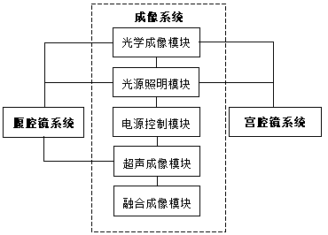
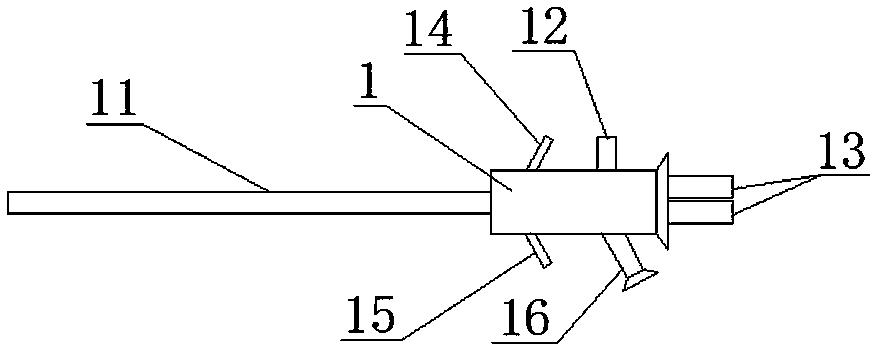
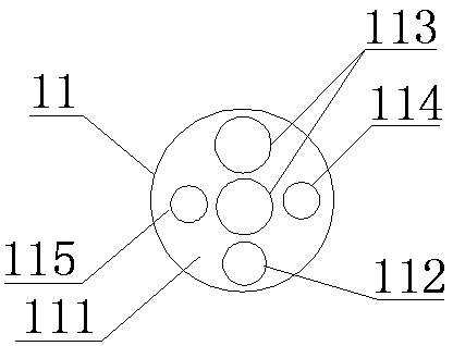
[0037] see figure 1 , an embodiment of the present invention provides a laparoscopic myomectomy system, including a laparoscopy system, a hysteroscopy system, and an imaging system; the imaging system includes a power control module, a light source lighting module, an optical imaging module, an ultrasonic An imaging module and a fusion imaging module, the power control module is respectively connected to the light source lighting module and the ultrasound imaging module, the optical imaging module is connected to the light source lighting module, and the fusion imaging module is connected to the ultrasound imaging module The modules are connected; the laparoscope system is connected with the light source lighting module, the optical imaging module and the ultrasonic imaging module respectively; the hysteroscope system is connected with the light source lighting module and the optical imaging module respectively, and the uterine cavity The endoscopic system includes resection i...


PUM


Abstract

The invention discloses a hysteromyoma excision system under a hysteroscope combined with a laparoscope. The system comprises a laparoscope system, a hysteroscope system and an imaging system. The laparoscope system is separately connected with a light source illumination module, an optical imaging module and an ultrasonic imaging module in the imaging system; the hysteroscope system is separatelyconnected with the light source illumination module and the optical imaging module in the imaging system and comprises an excision instrument for integrally excising the hysteromyoma. According to the hysteromyoma excision system under the laparoscope combined with the laparoscope, an ultrasonic probe is additionally arranged in the laparoscope, an ultrasonic image is fused with a preoperative magnetic resonance image, and therefore invisible myometrial myomas can be precisely imaged; the myomas under the hysteroscope are integrally excised so as to avoid the residues of myoma tissue fragments, and therefore the comprehensive excision of the hysteromyoma is realized; the hysteromyoma excision system has the advantages of convenient operation, small wounds, less bleeding, quick recovery, high myoma stripping rate and the like, can effectively reduce the residual rate and the relapse rate after an operation and has a good prognosis effect.

Description

technical field [0001] The invention relates to the technical field of medical devices, in particular to a laparoscopic myomectomy system. Background technique [0002] Uterine fibroids are common gynecological tumors, generally manifested as clinical symptoms such as uterine bleeding, abdominal mass, pain, and increased leucorrhea. If not treated in time, the tumor may deteriorate and seriously endanger the patient's physical and mental health. The traditional treatment method for uterine fibroids is laparotomy, but this method is risky and traumatic, and the postoperative recovery is slow, and it will have a certain impact on the fertility of the patient, so it is gradually being treated with less damage, faster recovery, and more impact on organs. Minor hysteroscopy and laparoscopy alternatives. At present, hysteroscopic surgery and laparoscopic surgery are two common surgical methods for hysteromyomectomy, which can be used alone or in combination. Multiple uterine fib...

Claims


Application Information

Patent Timeline
IPC IPC(8): A61B17/00A61B17/42A61B90/17A61B17/3205A61B34/20A61B18/12A61B18/14A61B34/10A61B1/303A61B1/313A61B1/04A61B1/07
CPCA61B1/04A61B1/07A61B1/303A61B1/3132A61B17/00234A61B17/320016A61B17/3205A61B17/4241A61B18/12A61B18/14A61B34/10A61B34/20A61B90/17A61B2017/00287A61B2017/4216A61B2018/00559A61B2018/00577A61B2018/00589A61B2034/107A61B2034/108A61B2034/2063A61B2034/2065
Inventor 颜磊张芳芳
Owner SUZHOU BASECARE MEDICAL DEVICE CO LTD
Learn More