
Surgical microimaging system based on optical coherence tomography augmented reality

A technology of optical coherence tomography and microscopic imaging, applied in surgical microscopes, surgical navigation systems, surgery, etc., can solve the problems of poor recognition effect, limited application, trauma, etc., and achieve the effect of intuitive surgical guidance

Active Publication Date: 2020-01-03
SUZHOU INST OF BIOMEDICAL ENG & TECH CHINESE ACADEMY OF SCI
View PDF6 Cites 11 Cited by
  • Summary
  • Abstract
  • Description
  • Claims
  • Application Information

AI Technical Summary

Problems solved by technology

The key to augmented reality technology lies in virtual-real registration, that is, to establish the coordinate transformation relationship between virtual images and real scenes. Registration as a reference point can get a matching result with better accuracy, but this method may be traumatic; using body surface features to set a reference point can avoid additional trauma, but the recognition effect is not good when the features are not obvious, limiting application of the method

Method used


Image

  • Surgical microimaging system based on optical coherence tomography augmented reality
  • Surgical microimaging system based on optical coherence tomography augmented reality
  • Surgical microimaging system based on optical coherence tomography augmented reality

Examples


Embodiment Construction

[0045] The present invention will be further described in detail below in conjunction with the embodiments, so that those skilled in the art can implement it with reference to the description.

[0046] It should be understood that terms such as "having", "comprising" and "including" used herein do not exclude the presence or addition of one or more other elements or combinations thereof.

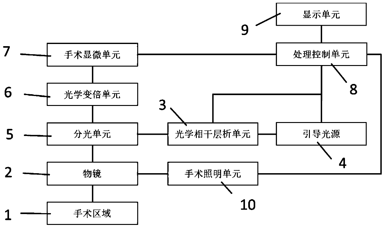
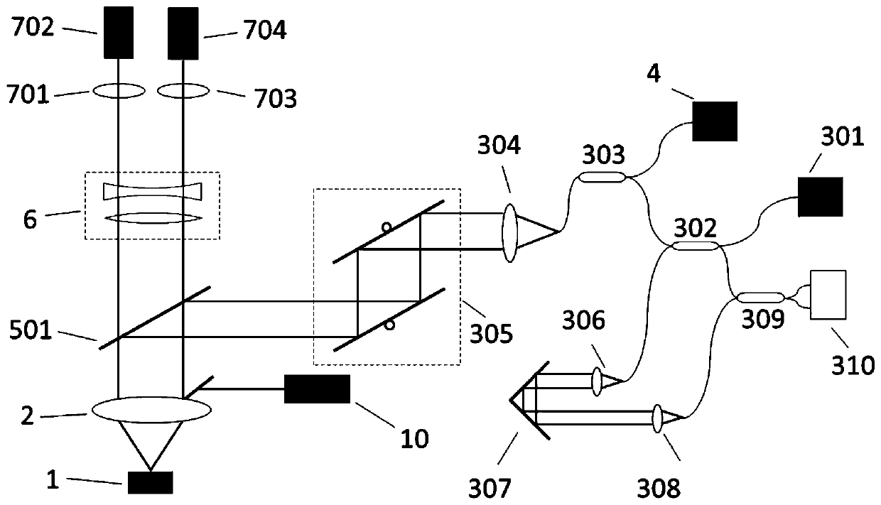
[0047] Such as Figure 1-2 As shown, a surgical microscopic imaging system based on optical coherence tomography augmented reality in this embodiment includes:

[0048] A surgical microscopic unit 7, configured to collect two-dimensional microscopic images of the surgical region 1;

[0049] The optical coherence tomography unit 3 is used to collect the OCT three-dimensional image of the operation area 1;

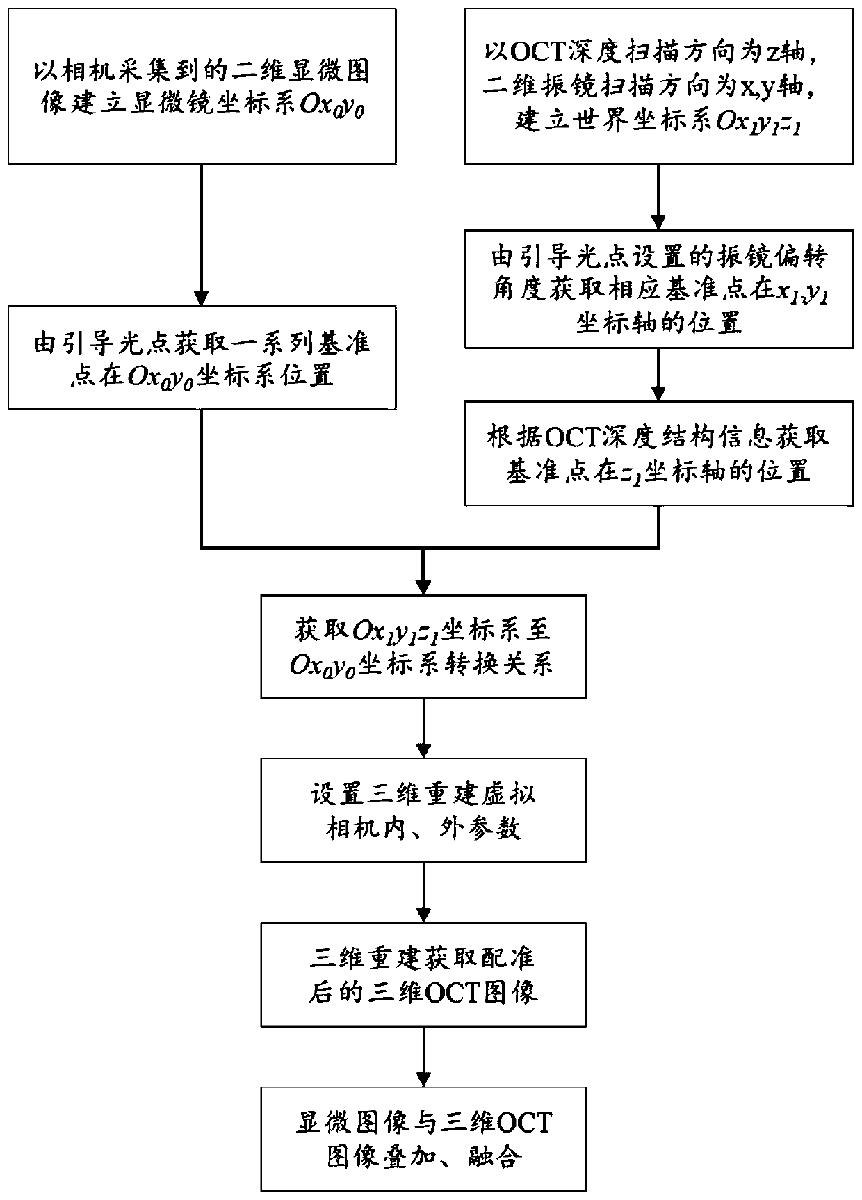
[0050] The guiding light source 4, which can be captured by the camera of the operating microscopic unit 7, is used to project the guiding light spot synchronized with the OCT scanning l...


PUM


Abstract

The invention discloses a surgical microimaging system and method based on optical coherence tomography augmented reality. The system comprises a surgical microscopic unit which is used for collectingtwo-dimensional microscopic images of a surgical region, an optical coherence tomography unit which is used for collecting OCT three-dimensional images of the surgical region, a guiding light sourcewhich can be captured by a camera of the surgical microscopic unit and is used for projecting a guiding light spot synchronized with an OCT scanning light source of the optical coherence tomography unit, a processing control unit which is used for acquiring the two-dimensional microscopic images and OCT three-dimensional images of the surgical region, as well as images after the two-dimensional microscopic images and the OCT three-dimensional images of the surgical region are fused, and a display unit which is used for outputting and displaying results of the control unit. The surgical microimaging system can be used for accurately registering and fusing two-dimensional microscopic images and OCT three-dimensional images, realizes microscopic image real-time augmentation of microscopic images of the surgical region, provides intuitive navigation information to surgery, and realizes intuitive surgical guidance.

Description

technical field [0001] The invention relates to the technical field of microsurgery imaging and graphic processing, in particular to a surgical microscopic imaging system based on optical coherence tomography augmented reality. Background technique [0002] Modern surgery has required that while locating the surgical target site, the physiological creation of the patient should be reduced as much as possible to achieve minimally invasive surgery. Image-guided interventional surgery can accurately locate the surgical target, can carry out preoperative planning before surgery, real-time monitoring and navigation during surgery, and evaluation of surgical effects in the surgical area after surgery. It has the advantages of high precision and small trauma. , is an important direction of modern surgery. [0003] At present, the imaging range of optical microscope-based microsurgery, such as ophthalmic surgery and neurosurgery, is limited to two-dimensional imaging of the surface...

Claims


Application Information

Patent Timeline
IPC IPC(8): A61B34/20A61B90/00
CPCA61B34/20A61B90/37A61B2090/3735A61B2034/2055A61B90/20G02B21/0012G02B21/365A61B2090/365A61B2090/367G06T19/006G06T2210/41G01B9/02091A61B3/102G01B9/0203G01B9/02004A61B90/30A61B2090/371G06T7/70H04N13/302H04N13/239G02B21/367G02B27/141G06T11/008G06T2207/10056G06T2207/10101G06F18/251H04N23/56H04N23/74
Inventor 史国华樊金宇何益邢利娜高峰
Owner SUZHOU INST OF BIOMEDICAL ENG & TECH CHINESE ACADEMY OF SCI
Learn More