
Reagent card for quantitatively detecting tumor necrosis factor alpha and preparation method thereof

A technology for quantitative detection of tumor necrosis factor, which is applied in the field of reagent card for quantitative detection of tumor necrosis factor alpha and its preparation, can solve the problems of many interference factors and quantitative determination, and achieves the effects of simple operation, time saving and fast detection speed.

Pending Publication Date: 2020-01-14
HEBEI GENSURE BIOTECH LIC
View PDF5 Cites 0 Cited by
  • Summary
  • Abstract
  • Description
  • Claims
  • Application Information

AI Technical Summary

Problems solved by technology

Immunochromatographic method has been unable to carry out accurate quantitative determination due to many interference factors

Method used


Image

  • Reagent card for quantitatively detecting tumor necrosis factor alpha and preparation method thereof
  • Reagent card for quantitatively detecting tumor necrosis factor alpha and preparation method thereof
  • Reagent card for quantitatively detecting tumor necrosis factor alpha and preparation method thereof

Examples


Embodiment 1

[0066] Example 1 A Reagent Card for Quantitative Detection of Tumor Necrosis Factor α

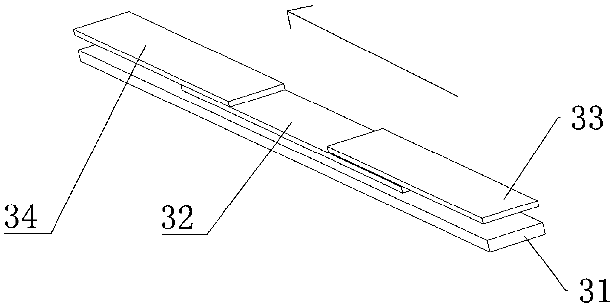
[0067] Such as figure 1 and figure 2 As shown, this embodiment includes a box body 1 and a reagent strip 3 arranged in the box body 1 . The reagent strip 3 includes a liner 31 on which an immunonitrocellulose membrane 32 is arranged. One end of the immunonitrocellulose membrane 32 is connected with an immunofluorescent material release pad 33 , and the other end is connected with an absorbent paper 34 .

[0068] The immunofluorescent material release pad 33 is coated with fluorescently labeled TNFα monoclonal antibody. A TNFα detection line 5 coated with a TNFα monoclonal antibody and a quality control line 4 coated with a goat anti-mouse IgG polyclonal antibody are arranged in parallel on the immunonitrocellulose membrane 32, and the TNFα detection line 5 is located at the quality control line 4 Between the release pad 33 and the immunofluorescent material;

[0069] A sampling hole 6 ...

Embodiment 2

[0073] Example 2 Preparation method of a reagent card for quantitative detection of tumor necrosis factor α

[0074] This embodiment is used for the preparation of Example 1, comprising the following steps carried out in sequence:

[0075] 1. Preparation of Immunofluorescence Material Release Pad 33

[0076] 1) Add labeling buffer to the fluorescent material and dilute the fluorescent material to VOL 1 =0.25%, obtain solution A;

[0077] Among them, the amount of fluorescent material = batch volume × target fluorescent material concentration 0.25% ÷ original fluorescent material concentration 1%, the amount of adding labeling buffer = batch volume - fluorescent material volume;

[0078] 2) Put solution A in n 1 = Centrifugal t at 12500rpm 1 = 15 minutes, discard the supernatant, redissolve the pellet with labeling buffer, sonicate, and then in n 2 = Centrifugal t at 12500rpm 2 = 15 minutes, discard the supernatant, add labeling buffer, and sonicate to obtain solution B; ...

Embodiment 3-6

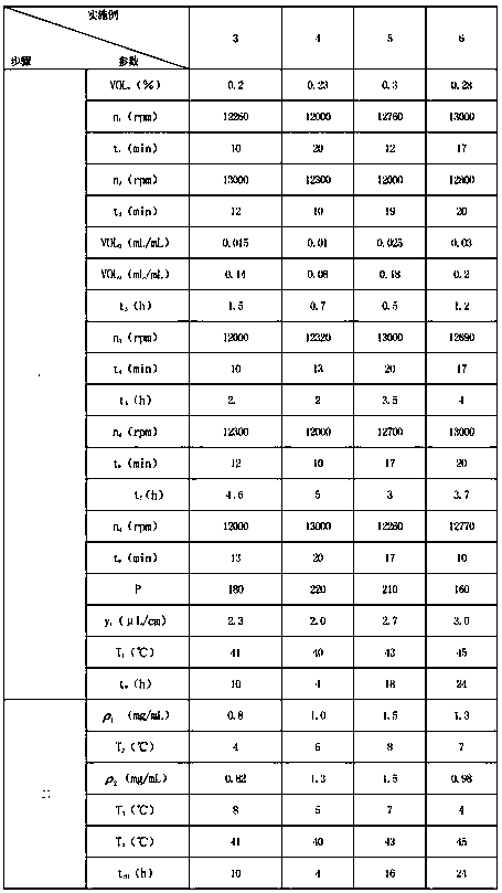
[0104] Example 3-6 Preparation method of a reagent card for quantitative detection of tumor necrosis factor α

[0105] The preparation process of embodiment 3-6 is substantially the same as embodiment 2, and the difference lies in the difference of parameters, and the specific parameters are as shown in table 1 below:

[0106] The parameter of table 1 embodiment 3-6

[0107]


PUM


Abstract

The invention discloses a reagent card for quantitatively detecting tumor necrosis factor alpha. The reagent card comprises a box body and a reagent strip arranged in the box body, wherein the reagentstrip comprises a pad, an immune nitrocellulose membrane is arranged on the pad, one end of the immune nitrocellulose membrane is linked with an immunofluorescence material release pad, and the otherend of the immune nitrocellulose membrane is linked with absorbent paper; the immunofluorescence material release pad is coated with a TNF alpha monoclonal antibody; the immune nitrocellulose membrane is provided with a TNF alpha detection line and a quality control line, and the TNF alpha detection line is positioned between the quality control line and the immunofluorescence material release pad; and the box body is provided with a sample adding hole and an observation window. The invention also discloses a preparation method of a kit for quantitatively detecting the tumor necrosis factor alpha. The kit adopts a dry-type fluorescence immunochromatographic technology and a double-antibody sandwich method principle to detect the concentration of the tumor necrosis factor alpha in a sample, can effectively eliminate the interference of non-specific fluorescence, improves the analysis sensitivity and the accuracy of quantitative determination, and is suitable for the technical field oftumor necrosis factor alpha detection.

Description

technical field [0001] The invention belongs to the field of biotechnology, and relates to a reagent card, in particular to a reagent card for quantitatively detecting tumor necrosis factor alpha and a preparation method thereof. Background technique [0002] Tumor necrosis factor is a cytokine with a wide range of biological activities. In 1985, Shalalby named the tumor necrosis factor produced by macrophages as tumor necrosis factor α. Studies have shown that tumor necrosis factor α is closely related to apoptosis. Some cells have death receptors on the surface, and death receptors sense cell cycle environment death signals , triggers the apoptosis mechanism in the cell through the binding of the ligand to the receptor. Tumor necrosis factor is a 17kDa non-glycosylated protein whose biologically active composition is a trimer. In addition to the soluble component of TNF-α, there is a membrane-bound component with a molecular weight of 17 kDa on the surface of TNF-produci...

Claims


Application Information

Patent Timeline
Patent Type & Authority Applications(China)
IPC IPC(8): G01N33/68G01N33/533
CPCG01N33/6863G01N33/533G01N2333/525
Inventor 陈庆全李伟甲
Owner HEBEI GENSURE BIOTECH LIC
Learn More