Primer pair and kit for rapidly detecting mycoplasma and application of primer pair and kit

A mycoplasma and primer pair technology, which is applied in biochemical equipment and methods, microorganisms, and microorganism-based methods, etc., can solve the problems of long culture time of mycoplasma and affect the therapeutic effect of cells, and achieve the effect of high sensitivity and quantitative detection.

Pending Publication Date: 2020-01-17
SHANGHAI EAST HOSPITAL EAST HOSPITAL TONGJI UNIV SCHOOL OF MEDICINE
View PDF6 Cites 0 Cited by
  • Summary
  • Abstract
  • Description
  • Claims
  • Application Information

AI Technical Summary

Problems solved by technology

In the "Chinese Pharmacopoeia", the methods for mycoplasma detection are mainly culture method and DNA staining method. As the gold standard detection method for mycoplasma detection, the culture method has high accuracy, but it usually takes 21 days to report the results, and the culture time is longer for slo

Method used


Image

  • Primer pair and kit for rapidly detecting mycoplasma and application of primer pair and kit
  • Primer pair and kit for rapidly detecting mycoplasma and application of primer pair and kit
  • Primer pair and kit for rapidly detecting mycoplasma and application of primer pair and kit

Examples


Embodiment 1

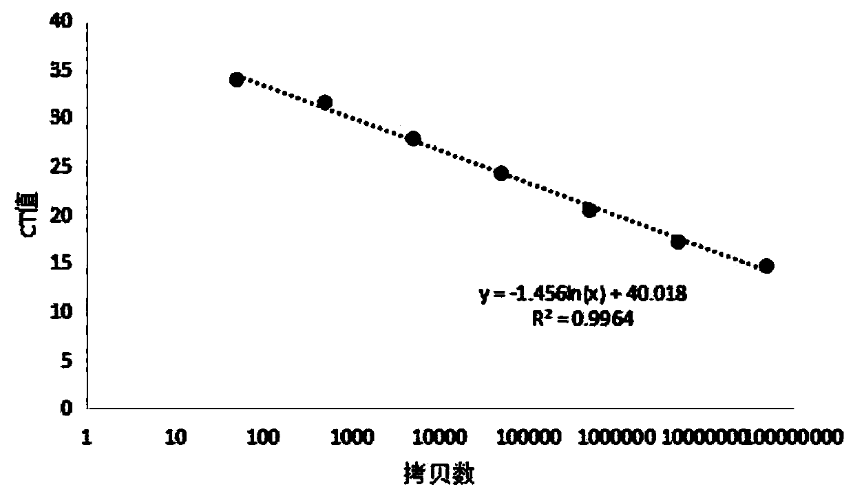
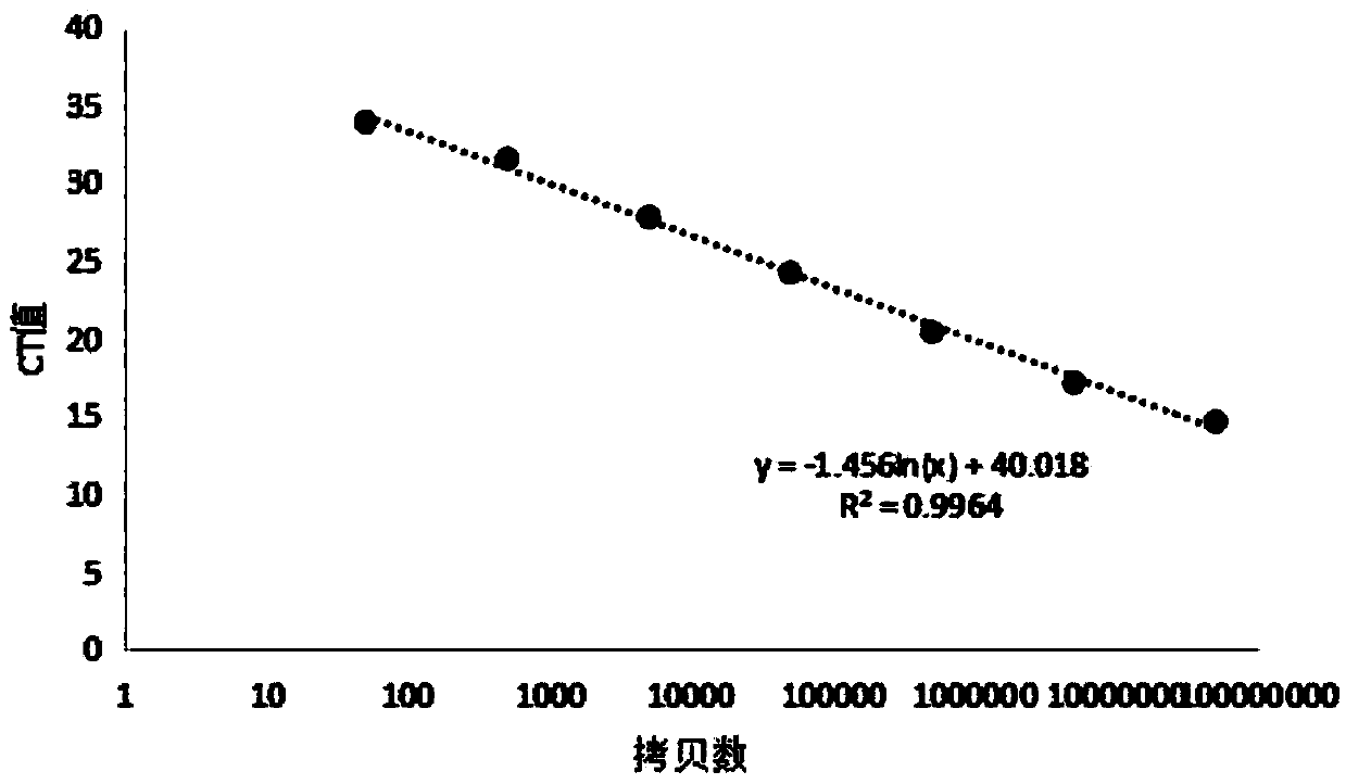
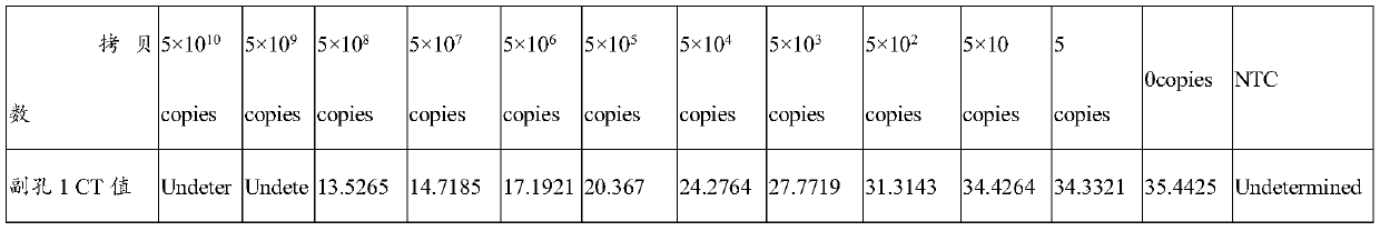
[0043] 1. Preparation of primers and standards

[0044] 1. Forward primer (SEQ ID No.1):

[0045] 5'-GAGCAAACAGGATTAGATACCCTGG-3',

[0046] or

[0047] Forward primer (SEQ ID No.2):

[0048] 5'-GAGCAAATAGGATTAGATACCCTGG-3';

[0049] Reverse primer (SEQ ID No.3):

[0050] 5'-CACCATCTGTCACTCTGTTAACCTCCA-3';

[0051] or

[0052] Reverse primer (SEQ ID No.4):

[0053] 5'-CACCATCTGTCACTCTGTTAACCTCCA-3'.

[0054] The primers were synthesized by Jerry Company, centrifuged at 12000g for 1min, each of the forward and reverse primers was dissolved in water to a concentration of 100μM, 2μl was added to 18μl of water and diluted to 10μM, and the remaining primers were stored at -20°C.

[0055] 2. The standard product is the pUC57 plasmid containing a partial sequence of the 16S rDNA conserved region, synthesized by Jerry Company. The base number of pUC57 is 2710bp. 16S rDNA partially conserved region sequence (SEQ ID No.3), base number 299bp. The standard was dissolved in 1×TEbuf...

Embodiment 2

[0066] Detection of human and mouse cells by QPCR

[0067] Detection of human and murine cells

[0068] Take cryopreserved human immune cells and rat stem cells, wash them with PBS for 3 times, then use Tiangen DNA Extraction Kit (DP304-02) to extract DNA, then take the DNA and use the present invention for detection, the detection method is the same as in Example 1 The results are shown in Table 2.

[0069] Table 2 Test results

[0070] sample concentration CT value tm human gene 61.7ng / μl 37.1 84.2℃ mouse gene 60.75ng / μl 37.6 84.2℃

[0071] It can be drawn from Table 2 that the amplification results of human and mouse genes using the primer pair of the present invention are negative, indicating that they are less affected by cell genes in cell detection.

Embodiment 3

[0073] Using the method of Example 1 to detect human umbilical cord mesenchymal stem cells and supernatant

[0074] After the human umbilical cord mesenchymal stem cells were grown to 80% confluence in the culture dish, 2ml of trypsin was added, digested at room temperature for 3min, and then culture medium was added to terminate the digestion.

[0075] After centrifugation at 100 g for 5 minutes, discard the supernatant, add 5 ml of normal saline, mix and wash three times, and take 100 μl of the added normal saline after centrifugation as test sample 1.

[0076] Add 100 μl of cell suspension to the bottom cells as test sample 2.

[0077] The sample to be tested was placed in a metal bath at 100° C. for 10 minutes, centrifuged at 12,000 g for 5 minutes, and 1 μl was added to the QPCR system in step 2 and step 2 of Example 1.

[0078] The positive control is the standard product diluted 2×10 4 times, the theoretical copy number is 2.5×10 6 , the negative control was water, a...


PUM


Abstract

The invention provides a primer pair and a kit for rapidly detecting mycoplasma and application of the primer pair and the kit and belongs to the technical field of mycoplasma detection. The primer pair comprises an upstream primer and a downstream primer; a nucleotide sequence of the upstream primer is shown as SEQ ID NO.1 or SEQ ID NO.2; and a nucleotide sequence of the downstream primer is shown as SEQ ID NO.3 or SEQ ID NO.4. The primer pair provided by the invention can specifically detect the mycoplasma, has no specificity to genes of human-origin cells and mouse-origin cells, can realizequantitative detection on the mycoplasma a limit of detection being 3.1*10-2.8-107 copies when being used for QPCR. The primer can realize rapid, extensive, high-sensitivity and quantifiable detection of the mycoplasma, is applied to the field of mycoplasma detection and is more suitable for mycoplasma detection in clinical research and application of cell and stem cell products.

Description

technical field [0001] The invention belongs to the technical field of mycoplasma detection, in particular to a primer pair and a kit for rapid detection of mycoplasma and applications thereof. Background technique [0002] Mycoplasma (Mycoplasma), also known as mycoplasma, is between bacteria and viruses in size. It is a class of smallest prokaryotic microorganisms that lack cell walls and can grow and reproduce in non-living media. Mycoplasma is simple in structure, small in size, highly pleomorphic, and can pass through bacteria filters (0.22-0.45 μm). It is widely distributed in nature, such as sewage, soil, ore, plants, insects, livestock and humans. It is susceptible to infection. Pathogens that cause various diseases in humans and animals are also pollutants that easily contaminate cells, strains of bacteria and chicken embryos used for production. In the "Chinese Pharmacopoeia", the methods for mycoplasma detection are mainly culture method and DNA staining method. ...

Claims


Application Information

Patent Timeline
IPC IPC(8): C12Q1/689C12Q1/04C12N15/11C12R1/35
CPCC12Q1/689
Inventor 刘中民贾文文白志慧汤红明
Owner SHANGHAI EAST HOSPITAL EAST HOSPITAL TONGJI UNIV SCHOOL OF MEDICINE
Why Eureka
Try Eureka