
A method for processing image data using a non-linear scaling model and a medical visual aid system

An image data and auxiliary system technology, applied in the field of endoscopy, can solve problems such as loss of information, saturation of image sensor pixels, etc., and achieve the effect of improving health monitoring

Pending Publication Date: 2020-02-07
安布股份有限公司
View PDF2 Cites 0 Cited by
  • Summary
  • Abstract
  • Description
  • Claims
  • Application Information

AI Technical Summary

Problems solved by technology

[0005] Although, this configuration is desirable due to the simplicity of the design, the light source may lead to overexposure of parts of the object to be viewed, thereby causing the pixels of the image sensor to saturate such that information about the object to be observed is lost

Method used


Image

  • A method for processing image data using a non-linear scaling model and a medical visual aid system
  • A method for processing image data using a non-linear scaling model and a medical visual aid system
  • A method for processing image data using a non-linear scaling model and a medical visual aid system

Examples


Embodiment Construction

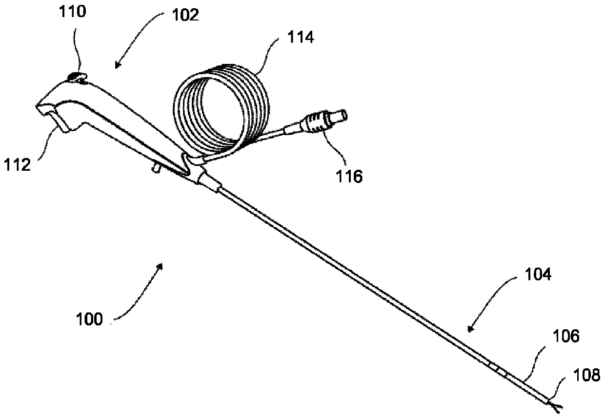
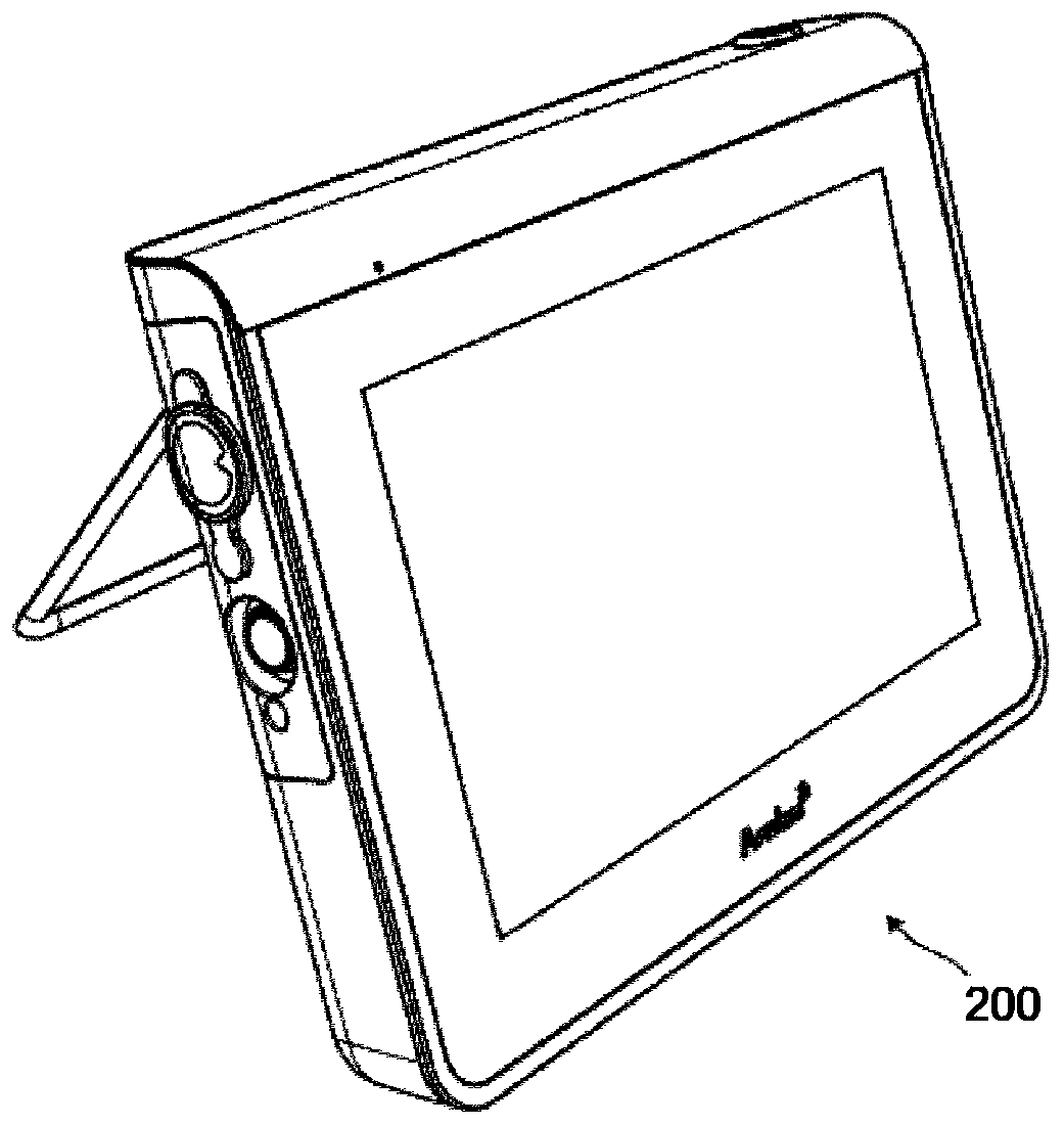
[0078] figure 1 An example of an endoscope 100 is shown. This endoscope may be suitable for single use. The endoscope 100 is provided with a handle 102 which is attached to an insertion tube 104 provided with a curved section 106 . The insertion tube 104 and the curved section 106 may be provided with one or several working channels, so that instruments such as holding devices can be inserted into the human body via the endoscope. One or more exit holes of the one or several channels may be provided in the tip portion 108 of the endoscope 100 . In addition to the exit aperture, a camera sensor, such as a CMOS sensor or any other image capture device, and one or several light sources, such as a Light Emitting Diode (LED) or any other light emitting device, may be placed in the tip section 108 . by making figure 2 The camera sensor and light source and monitor 200 shown in are configured to display images based on the image data captured by the camera sensor, the operator c...


PUM


Abstract

A method for obtaining and processing image data by using a medical visual aid system comprising an endoscope and a monitor is presented. The endoscope is configured to be inserted into a body cavityand comprises an image capturing device and a light emitting device. The method comprises illuminating a field of view of the image capturing device with the light emitting device, capturing the imagedata using the image capturing device, providing a non-linear scaling model adapted to the body cavity, adjusting the image data by applying the non-linear scaling model such that adjusted image datais formed, and presenting the adjusted image data on the monitor.

Description

technical field [0001] The present invention relates to endoscopes. More particularly, the invention relates to a method for obtaining and processing image data, a medical vision aid system, an endoscope forming part of a medical vision aid system, and a monitor forming part of a vision aid system. Background technique [0002] Endoscopes are well known devices for visual inspection of inaccessible places such as body cavities of the human body. Typically, an endoscope comprises an elongated insertion tube having a handle at its proximal end from the operator's point of view and an image sensor at its distal end, for example. Visual inspection devices such as built-in cameras and light sources. Endoscopes are usually connected to a monitor to display images captured by a camera when inserted into an object to be viewed. Wires for a camera and a light source (such as an LED) run along the interior of the elongated insertion tube from the handle to a tip at the distal end. ...

Claims


Application Information

Patent Timeline
IPC IPC(8): A61B1/00G06T5/00G06T5/10G06T5/40H04N5/235
CPCA61B1/00045A61B1/00057A61B1/00103G06T2207/10068A61B1/000095H04N23/56H04N23/80H04N23/63H04N23/74G06T5/90A61B1/0684G06T5/20G06T5/92
Inventor 芬恩·索尼伯格布瑞恩·尼尔森库尔特·阿兰·约翰松塞巴斯蒂安·奥尔特加·扎夫拉杨茜陈凯诚张玉旺
Owner 安布股份有限公司
Learn More