
Multiple-nuclide gamma imaging system and method

A nuclide and imaging technology, applied in the field of multi-nuclide gamma imaging systems, can solve the problems of difficulty in accurately delineating the scope of brain tumor tumor tissue infiltration, limited accuracy, and single information, and achieves wide practical value, The effect of improving the system signal-to-noise ratio and reducing the demand

Inactive Publication Date: 2020-02-11
NANCHANG UNIV
View PDF6 Cites 15 Cited by
  • Summary
  • Abstract
  • Description
  • Claims
  • Application Information

AI Technical Summary

Problems solved by technology

but single 18 F nuclide has the following disadvantages: First, because glucose is the main energy substance of tissues such as the brain, it normally takes high intake of 18F, and 18F is a non-specific positron tracer. First, some normal active tissues (such as cerebral cortex) The high uptake of 18F makes it difficult to accurately delineate the boundaries of brain tumors and the extent of tumor tissue infiltration on 18F-FDG PET imaging; 2. 18F-FDG is a non-specific tracer, and the increased uptake is not limited to malignant tumors, some benign tumors High uptake of 18F-FDG can also occur in lesions such as inflammation
These disadvantages of 18F nuclides on the gamma imaging system limit the accuracy of 18F-FDG PET imaging in the grading diagnosis of brain tumors to some extent
[0005] Existing PET gamma imaging devices are all single-nuclide imaging systems, which contain relatively simple information, while organisms are complex multi-molecular systems, and there are various forms and degrees of coupling between these multi-molecular nuclides , so it is urgently needed to provide more direct tumor cell information and precise treatment guidance for biological research and clinical medicine

Method used


Image

  • Multiple-nuclide gamma imaging system and method
  • Multiple-nuclide gamma imaging system and method
  • Multiple-nuclide gamma imaging system and method

Examples


Embodiment Construction

[0028] The technical solutions of the present invention will be further described below in conjunction with the accompanying drawings and through specific implementation methods.

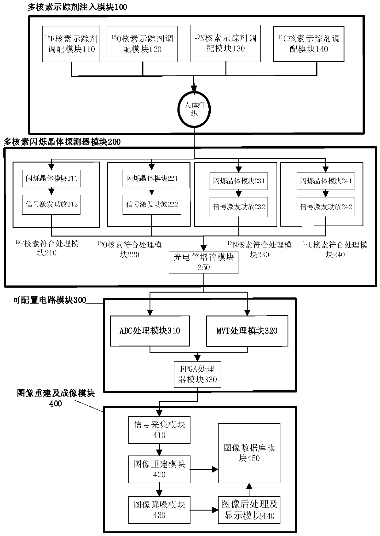
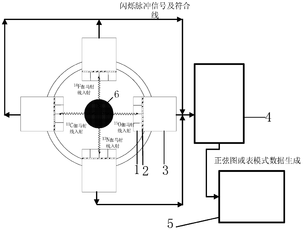
[0029] Such as Figure 1-Figure 4 As shown, according to one aspect of the present invention, a multi-nuclide gamma imaging system is provided in this embodiment, including a multi-nuclide tracer injection module 100, a multi-nuclide scintillation crystal detector module 200, which can Configuration circuit module 300, image reconstruction and imaging module 400, multi-nuclide tracer injection module 100 has a corresponding 11 C. 13 N. 15 O. 18 Four independently controllable channels of F gamma photon rays, the multi-nuclide scintillation crystal detector module 200 consists of corresponding 11 C. 13 N. 15 O. 18 F is made up of detectors of nuclides of four kinds of energies, which are fixed by a ring bracket, and an imaging object is placed in the center of the circle. The multi-nuclides tr...


PUM


Abstract

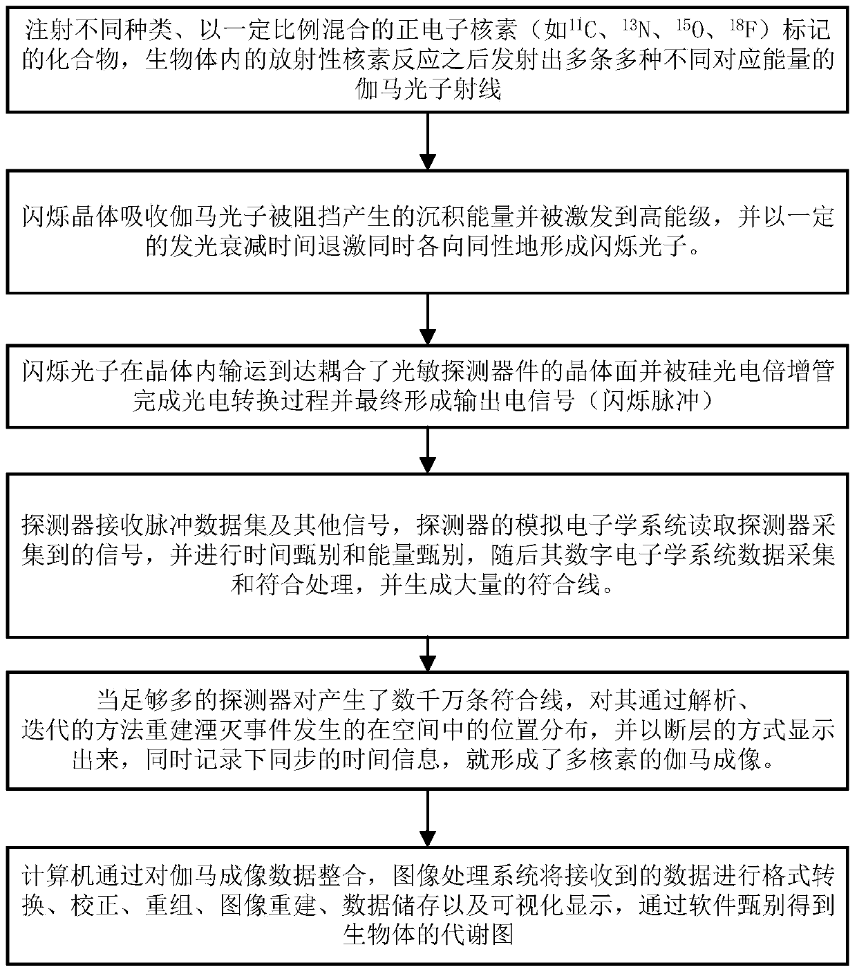
The present invention discloses a multiple-nuclide gamma imaging system and method and belongs to the field of medical imaging. The multiple-nuclide gamma imaging system comprises a multiple-nuclide tracer injection module, a multi-nuclide scintillation crystal detector module, a configurable circuit module and an image reconstruction and imaging module. The multiple-nuclide gamma imaging method comprises the following steps: S1, starting the imaging system, setting acquisition time of the imaging system, and injecting 11C, 13N, 15O and 18F labeled compounds into an imaging object; S2, obtaining a decay event emission gamma pulse data set by positioning a gamma detector; S3, outputting an electrical signal (scintillation pulse) by a silicon photomultiplier; S4, conducting time discrimination, energy discrimination, data acquisition and coincidence processing by a detector; and S5, displaying images in a tomographic manner by a computer. The disclosed multiple-nuclide gamma imaging system and method can obtain more comprehensive data than the prior art, comprehensively analyze multi-molecule events and related influences, reduce requirements on the total gamma photon counting, reduce irradiation risks on organisms and improve a signal-to-noise ratio of reconstructed images.

Description

technical field [0001] The invention relates to the field of medical imaging, in particular to a multi-nuclide gamma imaging system and method. Background technique [0002] Gamma or gamma-ray (γ-ray) imaging technology and systems play an important role in the research of many aspects of life science or biomedicine (such as genetics, genetics, etc.), especially in precision medicine and targeted diagnosis and treatment. It is an important means or approach for understanding, diagnosing, treating diseases and developing new drugs. It not only promotes the development of molecular biology and molecular medicine, but also promotes the progress of molecular imaging technology. It uses radionuclide-labeled tracer molecules to participate in the physiological metabolic process of organisms, detects X-rays or gamma photons emitted by radionuclide outside the body and reconstructs the image to obtain the distribution of radionuclide, so as to use radioactive elements Tracing metho...

Claims


Application Information

Patent Timeline
Patent Type & Authority Applications(China)
IPC IPC(8): A61B6/03
CPCA61B6/037A61B6/4258A61B6/5205A61B6/5211A61B6/5258
Inventor 邓贞宙陈冠东
Owner NANCHANG UNIV
Learn More