Microfluidic device and method for in-vitro 3D culture of cells and tissue

A microfluidic device and tissue culture technology, applied in tissue cell/virus culture devices, enzymology/microbiology devices, biochemical cleaning devices, etc., can solve the problem of different drug response modes and signal molecular pathways, and high experimental costs. , complex operation and other problems, to achieve the effect of efficient drug detection, short experimental period and easy operation

Pending Publication Date: 2020-03-27
ZHEJIANG UNIV
View PDF0 Cites 2 Cited by
  • Summary
  • Abstract
  • Description
  • Claims
  • Application Information

AI Technical Summary

Problems solved by technology

[0002] Two-dimensional cell culture lacks the source tissue microenvironment, which is significantly different from the growth mode of cells in vivo, resulting in different drug response modes and expression of signaling molecular pathways from those in vivo; in addition, animal models have long cycles, complicated operations, and high experimental costs. There are also some ethical issues
More importantly, the current culture device can only satisfy a single cell culture, and cannot be used for cell and tissue culture at the same time. Therefore, it is necessary to construct a device that can be used for cell and tissue culture in vitro to achieve More biomimetic cell and tissue culture
[0003] With the continuous development and progress of science and technology, there are many culture devices for cells and tissues, but most of the existing culture devices are limited to 2D culture of single cells, and it is impossible to perform cell and tissue culture on one device at the same time. bionic culture

Method used


Image

  • Microfluidic device and method for in-vitro 3D culture of cells and tissue
  • Microfluidic device and method for in-vitro 3D culture of cells and tissue
  • Microfluidic device and method for in-vitro 3D culture of cells and tissue

Examples


Embodiment 1

[0034] Example 1, a microfluidic culture device, referring to the attached Figure 1-2 .

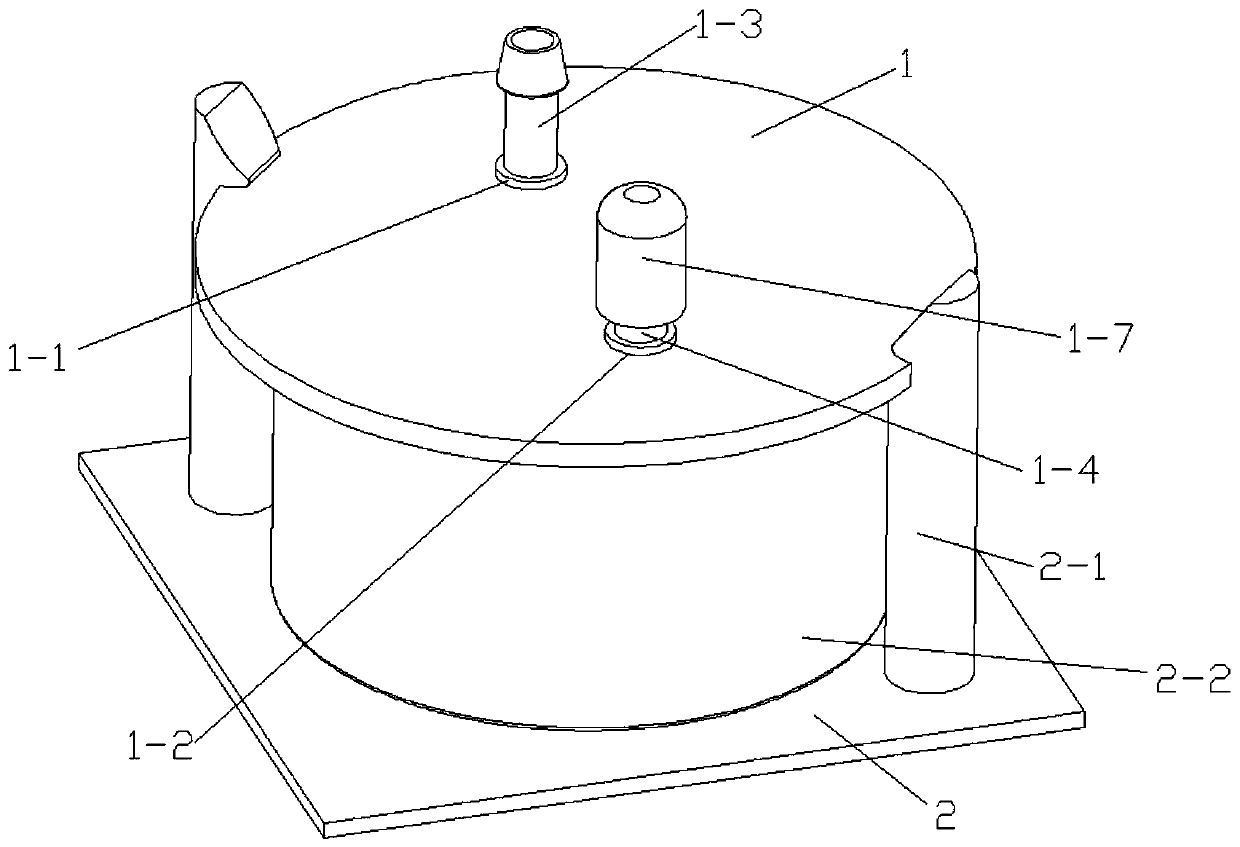
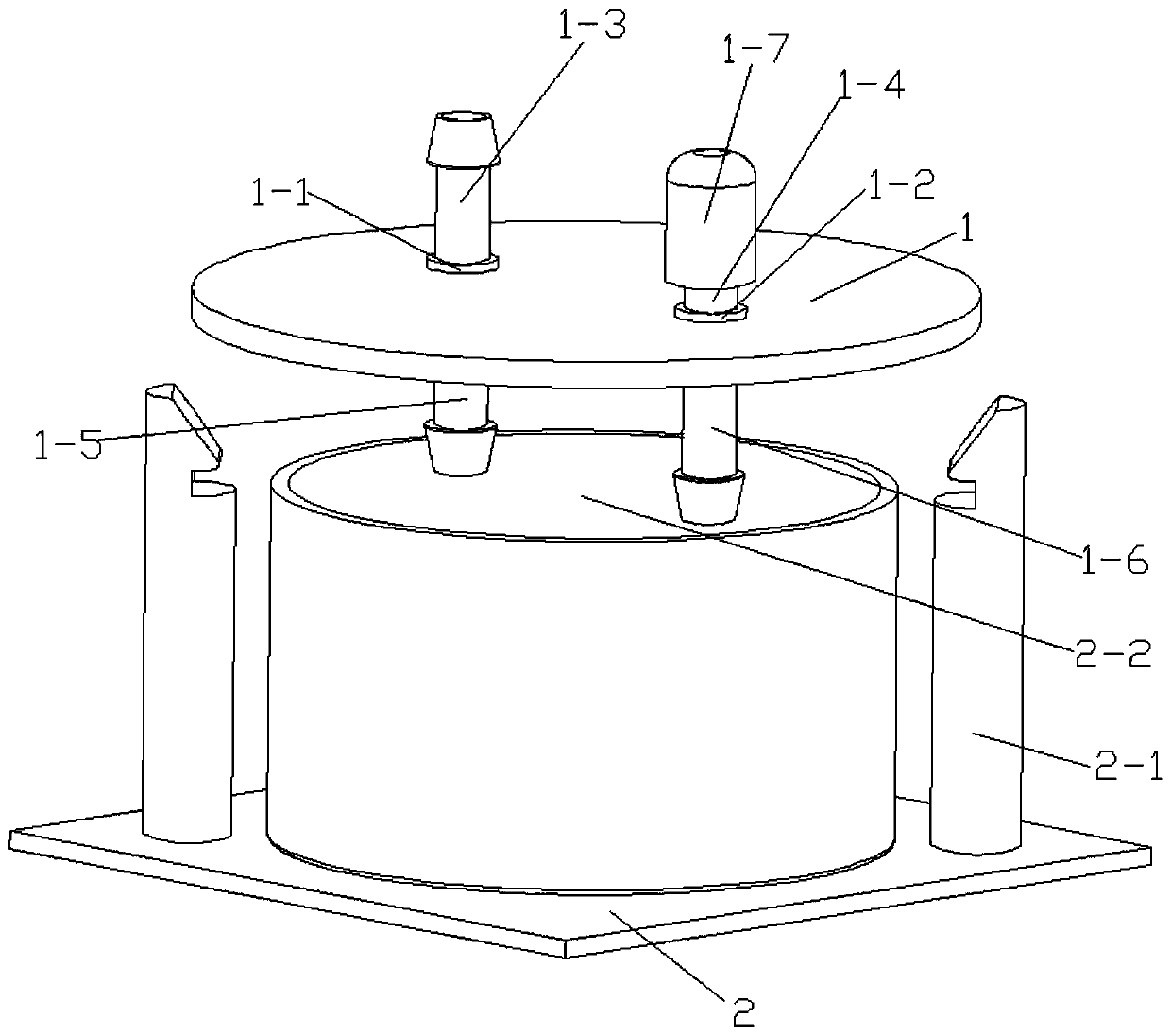
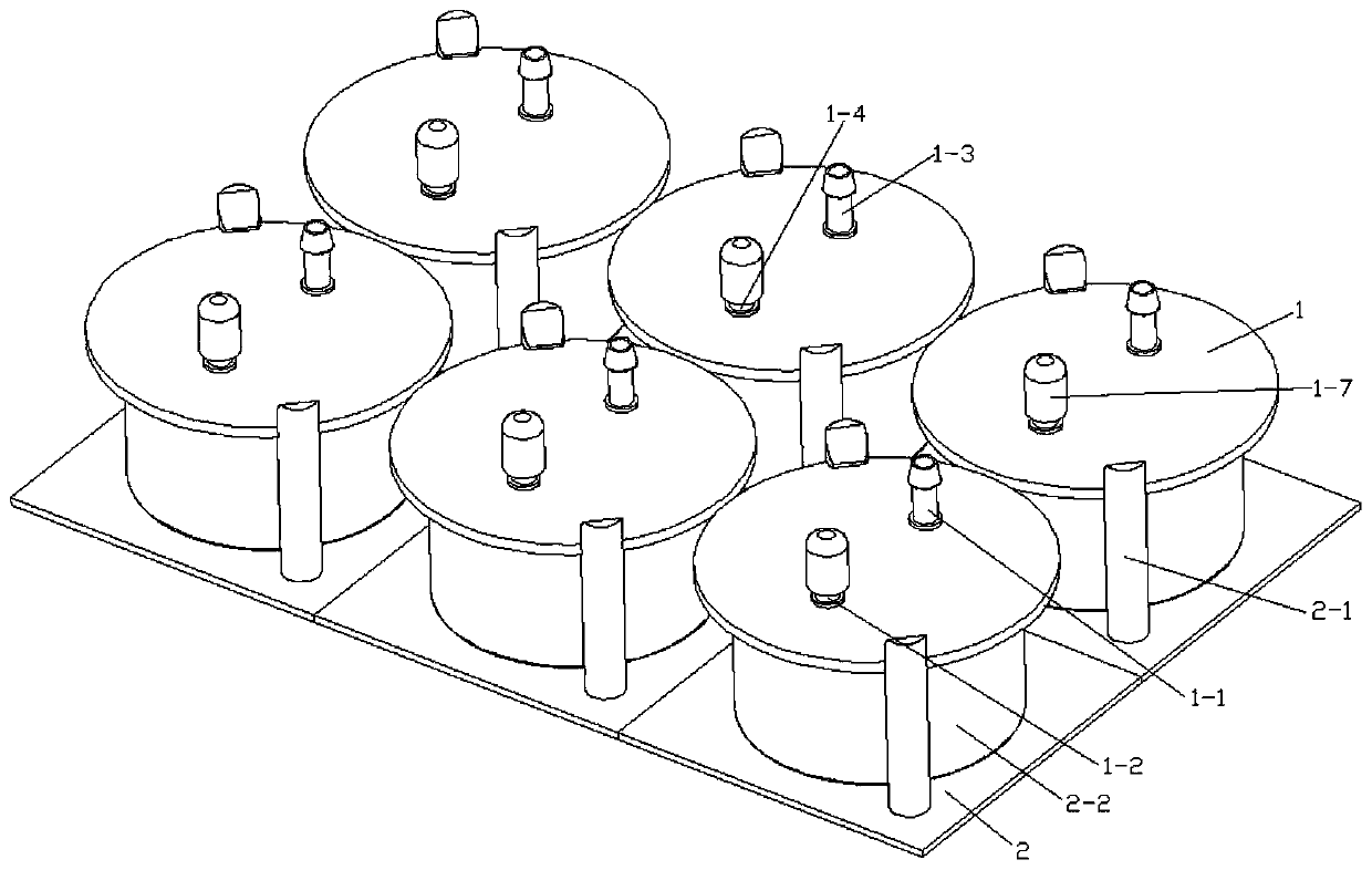
[0035] Such as figure 1 Shown is a culture unit in the overall device of the present invention. The culture unit can be used alone for cell or tissue culture, or can be connected in parallel or in series with other culture units on a peristaltic pump for cell or tissue culture.

[0036] The culture unit includes a cover layer 1, the cover layer 1 is provided with a liquid inlet hole 1-1 and a liquid outlet hole 1-2, the upper end of the liquid inlet hole 1-1 is provided with a first upper Luer connector 1-3, the liquid inlet The lower end of the hole 1-1 is provided with a first lower Luer connector 1-5, the upper end of the liquid outlet hole 1-2 is provided with a second upper Luer connector 1-4, and the lower end of the liquid outlet hole 1-2 is provided with a second The lower Luer connector 1-6, the first upper Luer connector 1-3, the first lower Luer connector 1-5, the second upp...

Embodiment 2

[0048] Example 2, a cell and tissue culture method using a microfluidic device, refer to the attached Figure 1-3 .

[0049] In this embodiment, a culture method for in vitro 3D cell and tissue culture is also disclosed. The method adopts the microfluidic device of Embodiment 1 and includes the following steps:

[0050] (1) In an environment with a temperature of 37°C, an oxygen concentration of 95%, and a carbon dioxide concentration of 5%, put the replanted decellularized liver into the culture pool 2-2 with tweezers, and use the holding device to hold the arm 2-1 , combine the cover layer 1, cell and tissue culture chamber layer 2 together.

[0051](2) The microfluidic device is externally connected to a peristaltic pump, and the culture medium is added at a speed of 3 mL / min for culturing. In the shown embodiment, the upper and lower Luer joints of the inlet hole 1-1 and the outlet hole 1-2 of the microfluidic device are connected with PE tubes, and the device is connect...


PUM

PropertyMeasurementUnit
Diameteraaaaaaaaaa
Heightaaaaaaaaaa
Diameteraaaaaaaaaa

Abstract

The invention provides a device and method for in-vitro 3D culture of cells and tissue. The device includes a cover layer and a cell-tissue culture chamber layer, wherein the cover layer includes a liquid inlet hole, a liquid outlet hole, Luer joints are arranged on upper parts and lower parts of the liquid inlet hole and the liquid outlet hole, and the Luer joints can be externally connected to PE tubes; and the cell-tissue culture chamber layer includes multiple cell-tissue organ culture tanks, and buckle arms are arranged on both sides of each culture tank. When the device is applied to cell culture, multiple cell-tissue culture experiments can be performed in vitro at the same time, and the culture state of cells and tissue can be detected and monitored conveniently and quickly throughthe microfluidic design.

Description

technical field [0001] The invention belongs to the field of biological materials, and in particular relates to a microfluidic device for 3D cell and tissue culture in vitro. Background technique [0002] Two-dimensional cell culture lacks the source tissue microenvironment, which is significantly different from the growth mode of cells in vivo, resulting in different drug response modes and expression of signaling molecular pathways from those in vivo; in addition, animal models have long cycles, complicated operations, and high experimental costs. At the same time, there are certain ethical issues. More importantly, the current culture device can only satisfy a single cell culture, and cannot be used for cell and tissue culture at the same time. Therefore, it is necessary to construct a device that can be used for cell and tissue culture in vitro to achieve More biomimetic cell and tissue culture. [0003] With the continuous development and progress of science and techn...

Claims


Application Information

Patent Timeline
IPC IPC(8): C12M3/00C12M1/00
CPCC12M23/16C12M21/08
Inventor 王书崎李玉峰武国华吴迪吴建国刘慧石胜宇
Owner ZHEJIANG UNIV
Why Eureka
Try Eureka