Ultrasonic tomography three-dimensional imaging method and system based on spiral scanning

A technology of three-dimensional imaging and helical scanning, which is applied in ultrasonic/sonic/infrasonic diagnosis, ultrasonic/sonic/infrasonic Permian technology, ultrasonic/sonic/infrasonic image/data processing, etc., and can solve problems such as difficulties in 3D reconstruction, Achieve the effect of shortening the overall scanning time, improving quality, and improving interlayer resolution

Active Publication Date: 2020-04-21
WUHAN WESEE MEDICAL IMAGING CO LTD
View PDF4 Cites 4 Cited by
  • Summary
  • Abstract
  • Description
  • Claims
  • Application Information

AI Technical Summary

Problems solved by technology

The system adopts a tomographic scanning mode similar to conventional X-tomography, and obtains information on discrete layers of tissue. Due to the influence of scanning time on the results, the interval between layers is at the millimeter level, while the resolution of ultrasonic tomographic images reaches Submillimeter level, which brings difficulties to subsequent 3D reconstruction

Method used


Image

  • Ultrasonic tomography three-dimensional imaging method and system based on spiral scanning
  • Ultrasonic tomography three-dimensional imaging method and system based on spiral scanning
  • Ultrasonic tomography three-dimensional imaging method and system based on spiral scanning

Examples


Embodiment 1

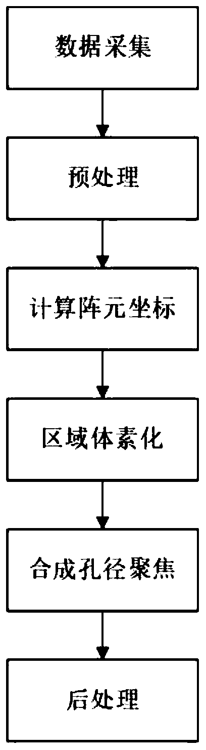
[0057] In this embodiment, the ultrasonic tomographic three-dimensional imaging method based on helical scanning specifically includes the following steps:

[0058] (1) Data collection:

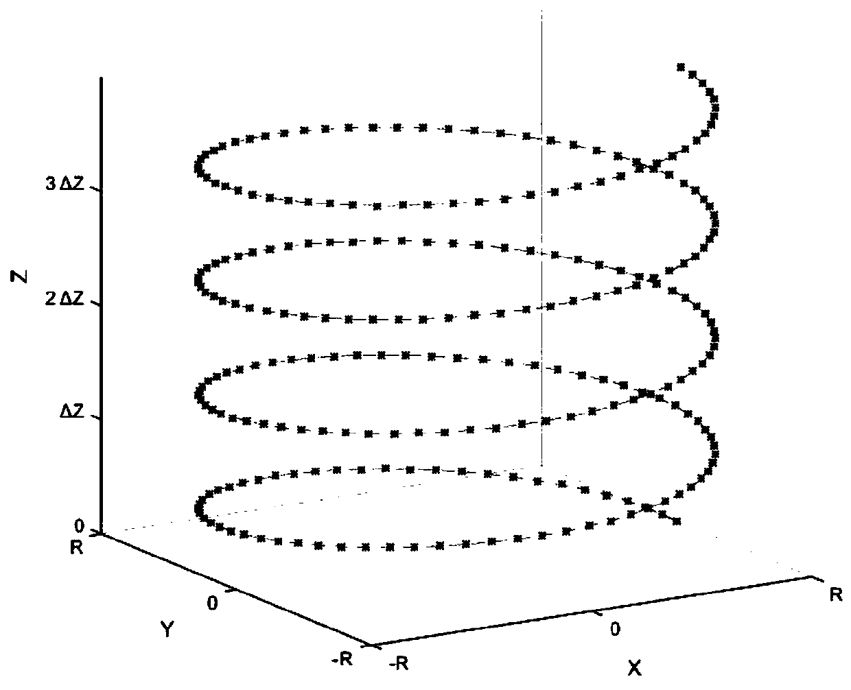
[0059] The uniformly distributed array elements on the circular detector are numbered from 1 to N, and the N is the number of array elements in the detector. A period of time before the start of acquisition, the detector is pulled by the motor to move at a uniform speed (defining that the direction of motion is parallel to The Z-axis direction of the three-dimensional direct coordinate system in space), and the speed is recorded as S; when the acquisition starts, array element 1 emits ultrasonic waves, and all N array elements receive them. After a time interval T, array element 2 emits ultrasonic waves, and all N array elements The reception of the array elements is carried out sequentially. When the transmission of the array element N is completed, the cycle is repeated to the array element...

Embodiment 2

[0092] In this embodiment, the ultrasonic tomographic three-dimensional imaging method based on helical scanning specifically includes the following steps:

[0093] (1) Data collection:

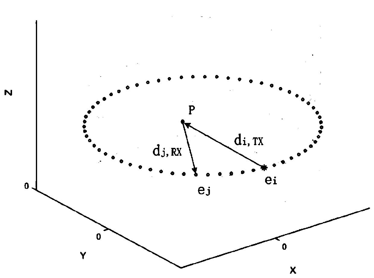
[0094] Assume that the angle of the center of the arc corresponding to the part of the circular detector is θ o , the radius of the arc is R, and the uniformly distributed array elements on some annular detectors are numbered from 1 to N, and the N is the number of array elements in the detector. Before starting to collect, the detector is pulled by the motor at a constant speed Movement (defining that the movement direction is parallel to the Z-axis direction of the three-dimensional direct coordinate system in space), and the speed is recorded as S; when the acquisition starts, the array element 1 emits ultrasonic waves, and all N array elements receive them. After the time interval T, the array element 2 Transmit ultrasonic waves, and all N array elements receive them sequentially. After ...

Embodiment 3

[0127] In this embodiment, the ultrasonic tomographic three-dimensional imaging method based on helical scanning specifically includes the following steps:

[0128] (1) Data collection:

[0129]The uniformly distributed array elements on the circular detector are numbered from 1 to N, and the N is the number of array elements in the detector. A period of time before the start of acquisition, the detector is pulled by the motor to move at a uniform speed (defining that the direction of motion is parallel to The Z-axis direction of the three-dimensional direct coordinate system in space), and the speed is recorded as S; when the acquisition starts, array element 1 and array element 2 emit ultrasonic waves at the same time, and all N array elements receive them. After a time interval T, array element 3 and array element 2 Unit 4 transmits ultrasonic waves, and all N array units receive them sequentially. When all the transmissions are completed, loop to array unit 1 and array uni...


PUM


Abstract

The invention belongs to the field of ultrasonic tomography, and discloses an ultrasonic tomography three-dimensional imaging method and system based on spiral scanning. The method comprises the following steps: (1) collecting original data, namely, while a probe keeps uniform linear motion, switching a transmitting array element to enable the change track of the position of an equivalent transmitting array element along with time in a three-dimensional space to be in a spiral shape or a partially spiral shape, and receiving echo data; (2) carrying out data preprocessing; (3) calculating the coordinates of each equivalent transmitting array element; (4) calculating the coordinates of imaging focus points; (5) performing synthetic aperture focusing on each imaging focus point; and (6) carrying out data post-processing. The imaging method principle, the overall process design and the like are improved, volume data containing tissue continuous layer information is obtained in a spiral scanning mode, it is proposed for the first time that the synthetic aperture focusing technology is applied to the three-dimensional space, the resolution between layers can be improved, the scanning time is shortened, and the imaging quality of the system can be improved.

Description

technical field [0001] The invention belongs to the field of ultrasonic tomographic imaging, and more particularly relates to a method and system for ultrasonic tomographic three-dimensional imaging based on helical scanning. Background technique [0002] Ultrasound tomography has the advantages of non-invasiveness, no radiation, high resolution and high sensitivity, and has broad application prospects in the early diagnosis of soft tissue tumors, and has gradually become one of the hot spots in the field of ultrasound applications. [0003] Ultrasound tomography can be divided into two modes: reflection imaging and transmission imaging, wherein transmission imaging includes sound velocity reconstruction and attenuation reconstruction. The reflection map shows the structural information of the target, and the sound velocity map and attenuation map show the functional information of the target. The ultrasonic tomography system uses step-by-step scanning to obtain information...

Claims


Application Information

Patent Timeline
Patent Type & Authority Applications(China)
IPC IPC(8): A61B8/14A61B8/00A61B8/08
CPCA61B8/145A61B8/483A61B8/0825A61B8/085A61B8/4411A61B8/4488A61B8/5215G01S15/8927G01S15/8993A61B8/0866A61B8/5207A61B8/5223G01S15/8997G01S15/8945G01S15/892
Inventor 尉迟明丁明跃刘昭辉张求德宋俊杰王珊珊周亮刘阔林
Owner WUHAN WESEE MEDICAL IMAGING CO LTD
Why Eureka
Try Eureka