Antigen detection kit, preparation method for antigen detection kit, and antigen detection method
An antigen detection and kit technology, applied in the field of biochemistry, can solve problems such as inaccurate antigen detection results, and achieve the effects of avoiding steric hindrance effects, accurately determining antigen content, and ensuring binding rate.
- Summary
- Abstract
- Description
- Claims
- Application Information
AI Technical Summary
Problems solved by technology
Method used
Image
Examples
preparation example Construction
[0035] The embodiment of the present invention also provides a method for preparing an antigen detection kit, which includes the following steps: Step S102, adding biotin to the monoclonal antibody solution containing only Fab fragments for reaction.
[0036] Step S104, after adding phosphate buffer, perform ultrafiltration to remove free biotin in the solution.
[0037] Step S106, after concentration by ultrafiltration, dilute with a buffer matrix, and add monoclonal antibodies containing Fab fragments and Fc fragments after dilution to obtain R1 solution.
[0038] Step S108, adding the chemiluminescence label to the secondary antibody solution for reaction.
[0039] Step S110, after adding phosphate buffer, ultrafiltration to remove free chemiluminescent markers in the solution.
[0040] Step S112, after concentration by ultrafiltration, use buffer matrix to dilute, add streptavidin magnetic beads to obtain R2 solution.
[0041] In the embodiment of the present invention, ...
Embodiment 1
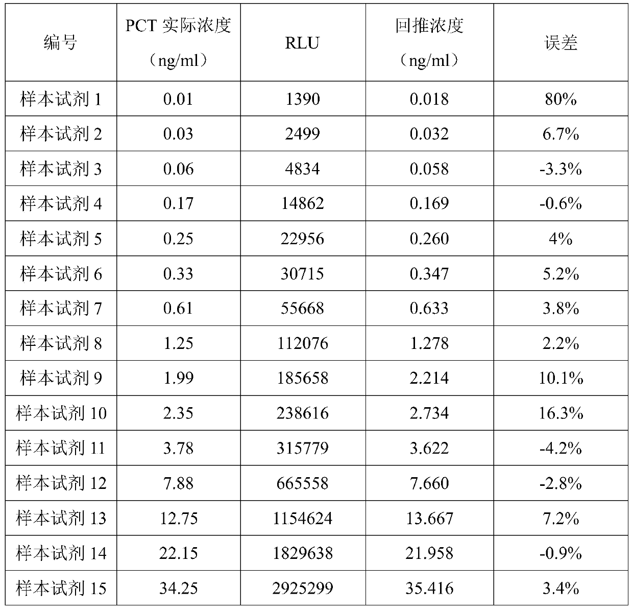
[0066] (1) Preparation of procalcitonin (PCT, PCT is used instead of procalcitonin) detection kit:
[0067] Add 1mg of biotin-NHS to 20mg of rabbit anti-human PCT monoclonal antibody, and incubate at 37°C for 30 minutes;
[0068] Use a 10mM / L phosphate buffer with a pH value of 7.0 to remove free biotin by ultrafiltration, and after ultrafiltration and concentration, use a buffer matrix to dilute to a biotin-labeled rabbit anti-human PCT monoclonal antibody concentration of 1 μg / mL. Buffer matrix includes phosphate buffer 10mM / L, bovine serum albumin 10g / L, Tween-20 5g / L, sodium chloride 8g / L and sodium azide 0.2g / L;
[0069] Add mouse anti-human PCT monoclonal antibody to the diluted buffer matrix until the concentration of mouse anti-human PCT monoclonal antibody is 1 μ g / mL to prepare R1 solution;
[0070] Add 1 mg of acridinium ester-NHS to 20 mg of goat anti-mouse IgG secondary antibody and incubate at 37°C for 30 minutes;
[0071] Use 10mM / L phosphate buffer with a pH ...
Embodiment 2
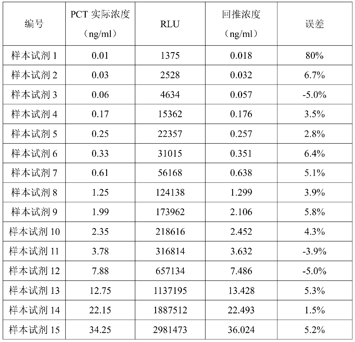
[0087] Identical to other steps of Example 1, the difference is only that (1) the steps of preparation of the procalcitonin detection kit are specifically:
[0088] Add 1 mg of biotin-NHS to 20 mg of rabbit anti-human PCT monoclonal antibody, and incubate at 37°C for 120 minutes;
[0089] Use a 10mM / L phosphate buffer with a pH value of 7.0 to remove free biotin by ultrafiltration, and after ultrafiltration and concentration, use a buffer matrix to dilute to a biotin-labeled rabbit anti-human PCT monoclonal antibody concentration of 100 μg / mL. Buffer matrix includes phosphate buffer 10mM / L, bovine serum albumin 10g / L, Tween-20 5g / L, sodium chloride 8g / L and sodium azide 0.2g / L;
[0090] Add mouse anti-human PCT monoclonal antibody to the diluted buffer matrix until the concentration of mouse anti-human PCT monoclonal antibody is 100 μ g / mL to prepare R1 solution;
[0091] Add 1 mg of acridinium ester-NHS to 20 mg of goat anti-mouse IgG secondary antibody and incubate at 37°C ...
PUM
| Property | Measurement | Unit |
|---|---|---|
| Concentration | aaaaa | aaaaa |
| Sensitivity | aaaaa | aaaaa |
Abstract
Description
Claims
Application Information
 - R&D
- Intellectual Property
- Life Sciences
- Materials
- Tech Scout
- Unparalleled Data Quality
- Higher Quality Content
- 60% Fewer Hallucinations
Browse by: Latest US Patents, China's latest patents, Technical Efficacy Thesaurus, Application Domain, Technology Topic, Popular Technical Reports.
© 2025 PatSnap. All rights reserved.Legal|Privacy policy|Modern Slavery Act Transparency Statement|Sitemap|About US| Contact US: help@patsnap.com


