3D printing distal tibial plate and preparation method therefor

A 3D printing, tibial technology, applied in the direction of outer plate, internal bone synthesis, medical science, etc., can solve the problems of personalized anatomical matching, insufficient fixation stability, etc., to reduce the risk of skin flap necrosis, high degree of anatomical matching, notch low effect

Inactive Publication Date: 2020-05-12
SOUTHERN MEDICAL UNIVERSITY
View PDF4 Cites 2 Cited by
  • Summary
  • Abstract
  • Description
  • Claims
  • Application Information

AI Technical Summary

Problems solved by technology

The preparation method of the 3D printed distal tibial plate solves the problems of personalized anatomical matching and insufficient fixation stability that are difficult to achieve with traditional tibial internal fixation plates

Method used


Image

  • 3D printing distal tibial plate and preparation method therefor
  • 3D printing distal tibial plate and preparation method therefor
  • 3D printing distal tibial plate and preparation method therefor

Examples


Embodiment 1

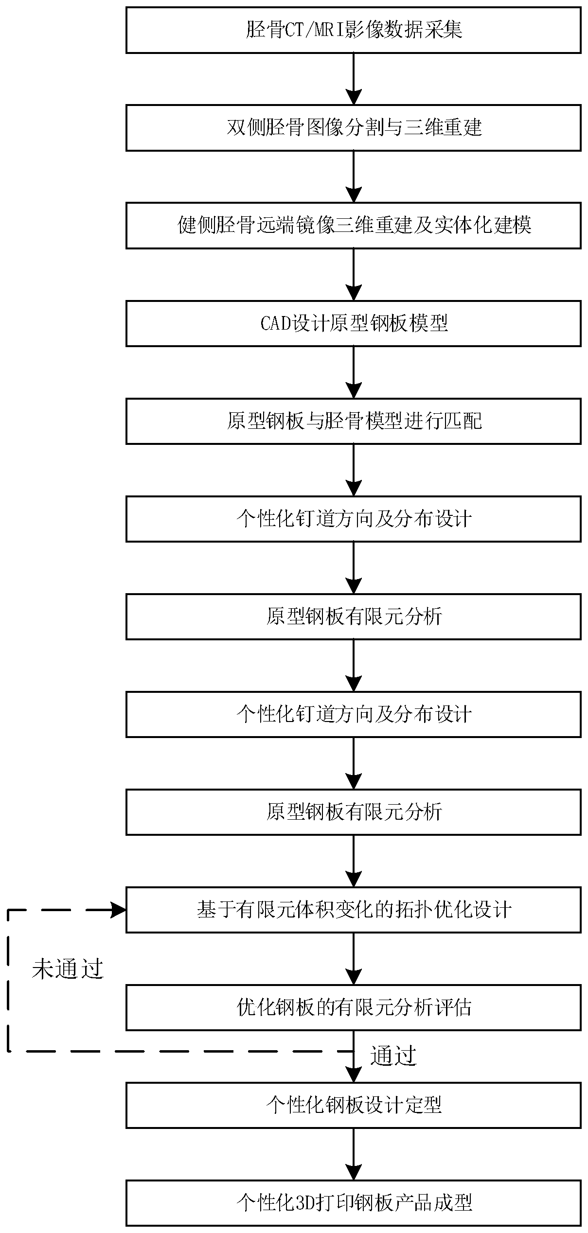
[0048] A method for preparing a 3D printed distal tibial plate, such as figure 1 As shown, the design method is realized through the following steps:

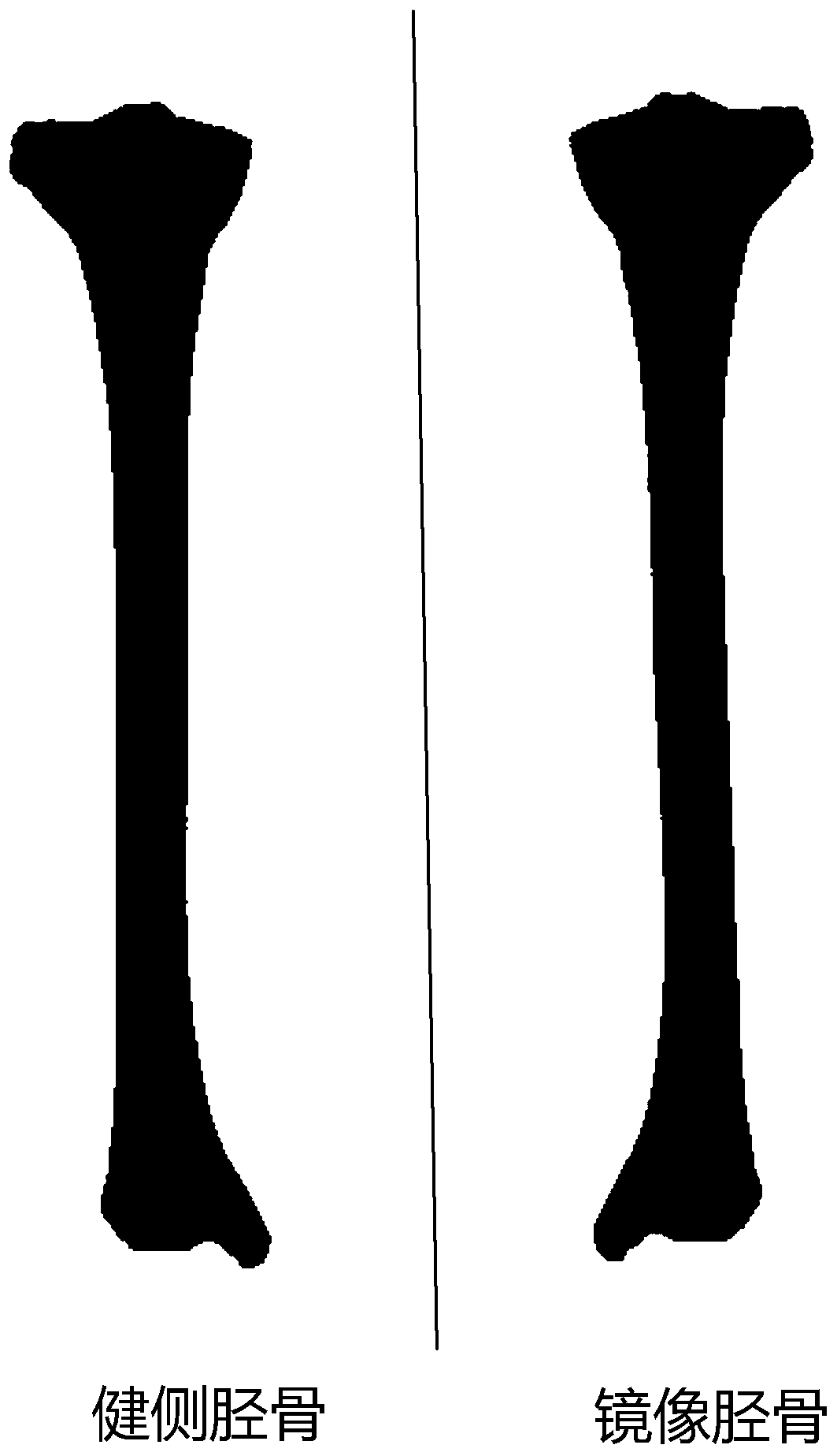
[0049] 1.1. Collect the computed tomography data of the affected side and the healthy bilateral tibia of the target object, and import the obtained computed tomography image data of the target object into the 3D reconstruction system. In the segmentation process, the initial three-dimensional model of the bilateral tibia of the target object is established, including the initial three-dimensional model of the healthy-side tibia and the initial three-dimensional model of the affected-side tibia of the target object.

[0050] Specifically, the computed tomographic image data of the bilateral tibia of the target object is collected, and the obtained computed tomographic image data of the bilateral tibia of the target object is imported into 3D reconstruction software for image segmentation processing, and a 3D model of the bilater...

Embodiment 2

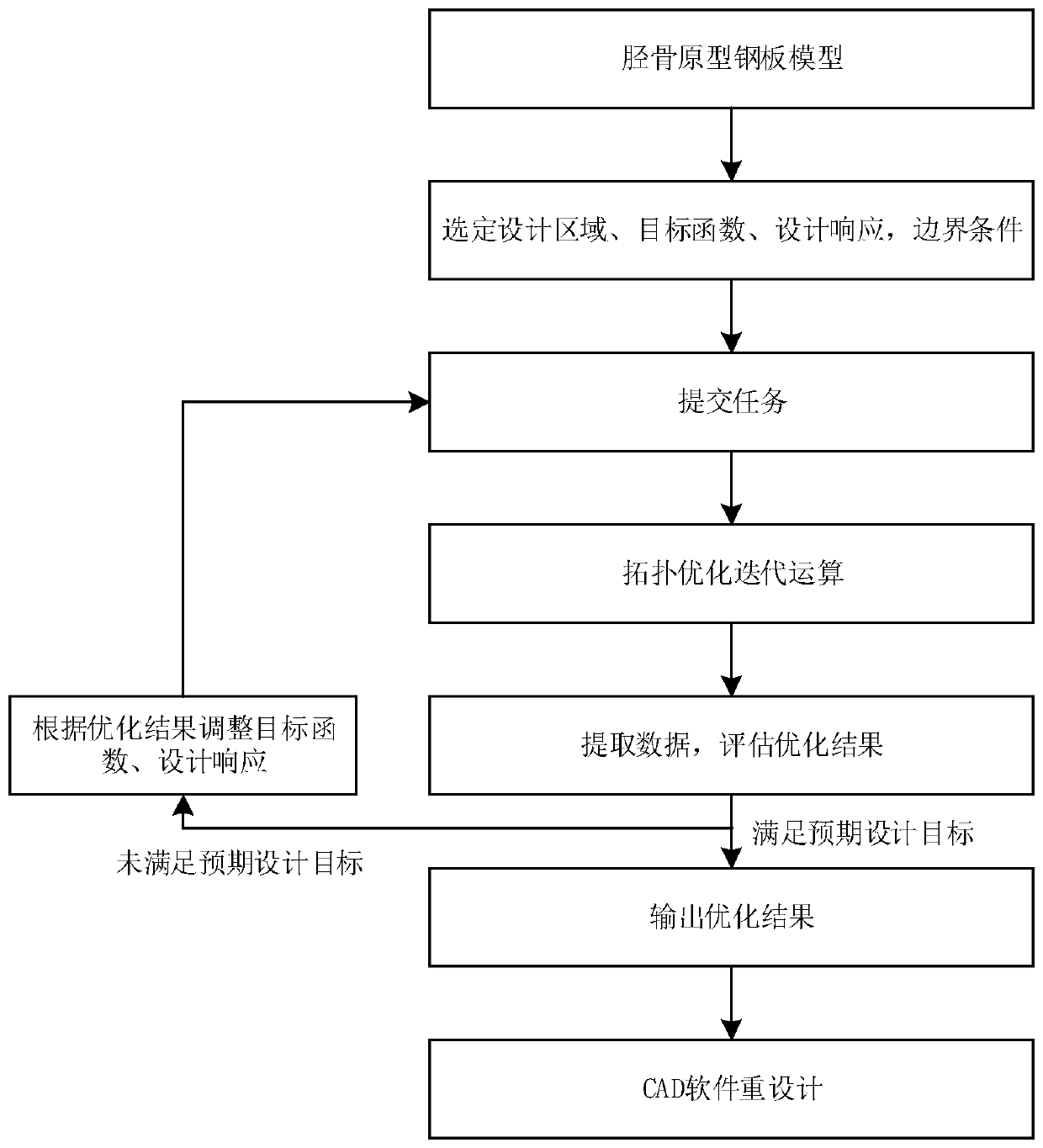
[0085] A method for preparing a 3D printed tibial plate, the other features of which are the same as those in Embodiment 1, except that the 3D printed tibial plate adopts the topology optimization technology of the finite element model.

[0086] The shape design of the steel plate with maximum volume reduction obtained by the topology optimization technology in the present invention can effectively reduce the notch of the steel plate on the premise of ensuring the maximum fixation rigidity, effectively protect the blood supply of local soft tissues and provide effective support for the collapsed articular surface , so as to avoid the related complications of autologous bone support and bone grafting, so that the tibia can fully restore its pre-injury shape.

Embodiment 3

[0088] A method for preparing a 3D printed distal tibia plate, which has the functional devices of Embodiment 1 and Embodiment 2. The difference is that the 3D printed tibial plate has a highly irregular and personalized surface,

[0089] Highly irregular and personalized curved surface, traditional machine tool processing is difficult to complete the forming and manufacturing of the steel plate through the cutting process, and the tibia steel plate designed by the present invention is directly manufactured by 3D printing forming method, realizing the direct forming of the steel plate, saving time and cost at the same time It also overcomes the shortcomings of traditional manufacturing techniques such as high cost and cumbersome procedures. At the same time, the personalized steel plate formed directly by 3D printing also avoids the decrease in mechanical strength caused by repeated shaping during the operation, and also greatly shortens the operation time.


PUM


Abstract

The invention relates to a preparation method for a 3D printing distal tibial plate. Computer tomography data of bilateral tibiae of the affected side and the healthy side of a target object is collected, the computer tomography data is utilized to establish a reference 3D model of the tibia of the affected side of the target object before fracture, and a tibial solid model is formed by fitting; the tibial solid model formed by fitting is imported into computer-aided design (CAD) to prepare a distal tibial prototype plate, then, finite element analysis and topological optimization are performed on the distal tibial prototype plate for the first time, and a model obtained after finite element analysis and topological optimization is imported into the CAD again to generate an optimized platemodel; finite element analysis and topological optimization are performed on the optimized plate model for the second time, whether a design expected standard is reached or not is judged, and if theexpected standard is reached, 3D printing preparation is performed, otherwise model optimization is performed again; and finally, the tibial plate is further processed. The method solves the problemsthat a traditional tibial internal fixation plate is hard to reach individualized anatomical matching, is insufficient in fixation stability, and the like.

Description

technical field [0001] The invention relates to the technical field of clinical repair and reconstruction of bone and joint injuries and the design of implantable medical devices, in particular to a 3D printed distal tibial plate and a preparation method thereof. Background technique [0002] Tibial fractures are a common type of extremity fractures, and distal tibial fractures account for about 7.2% of tibial fractures. Fractures of the distal tibia are often the result of high-energy injuries. At present, open reduction and internal fixation, as an irreplaceable treatment method for distal tibial fractures, is applied to fractures involving the articular surface that require anatomical reduction and unstable fractures in the distal 1 / 3 that require stability. [0003] Locking compression plates are commonly used clinically to fix distal tibial fractures. For those involving the articular surface of the distal tibia, multiple plates combined with multiple approaches may be...

Claims


Application Information

Patent Timeline
Patent Type & Authority Applications(China)
IPC IPC(8): A61B17/80A61B17/56B33Y50/00B33Y80/00
CPCA61B17/56A61B17/8061A61B2017/568B33Y50/00B33Y80/00
Inventor 黄文华王勉谭晋川邓羽平谢普生杨洋欧阳汉斌
Owner SOUTHERN MEDICAL UNIVERSITY
Why Eureka
Try Eureka