Non-local mean-guided partial volume correction method for PET images constrained by MR structural information

A non-local mean, partial volume technology, applied in the field of medical image processing, can solve the problems of Gibbs artifact, anatomical structure dependence, high level noise, etc., to improve accuracy and efficiency, improve accuracy, and improve PET images quality effect

Active Publication Date: 2021-03-09
SOUTHERN MEDICAL UNIVERSITY
View PDF12 Cites 0 Cited by
  • Summary
  • Abstract
  • Description
  • Claims
  • Application Information

AI Technical Summary

Problems solved by technology

By performing iterative deconvolution processing on the PET image itself, each voxel of the image can be corrected, but the correction process will introduce a high level of noise
In order to suppress the increase of noise, the existing median prior and wavelet filter-guided iterative deconvolution methods have achieved such an effect, but these deconvolution algorithms will have Gibbs artifacts
The PET partial volume correction method based on anatomical prior guidance has attracted widespread attention. However, the existing anatomical prior guided partial volume correction methods such as the RBV method have the problem of being too dependent on anatomical structures, tiny segmentation or registration. changes can lead to large differences in the results

Method used


Image

  • Non-local mean-guided partial volume correction method for PET images constrained by MR structural information
  • Non-local mean-guided partial volume correction method for PET images constrained by MR structural information
  • Non-local mean-guided partial volume correction method for PET images constrained by MR structural information

Examples


Embodiment 1

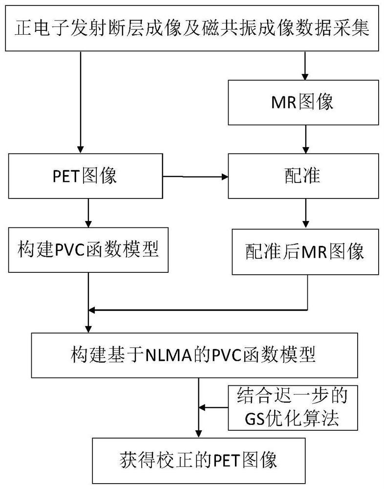
[0048] A non-local mean-guided partial volume correction method for PET images constrained by MR structural information, such as figure 1 As shown, the specific steps are:

[0049] Step (1) Synchronously collect PET and MR image data of the same object through PET equipment and MRI equipment respectively, obtain the PET image and MR image of the object synchronously, and obtain the system resolution of the detector in the PET imaging equipment at the same time;

[0050] Step (2) according to the PET image data that step (1) obtains, construct the model based on the partial volume correction of PET image under the PWLS framework;

[0051] Step (3) registering the MR image obtained in step (1) and the PET image;

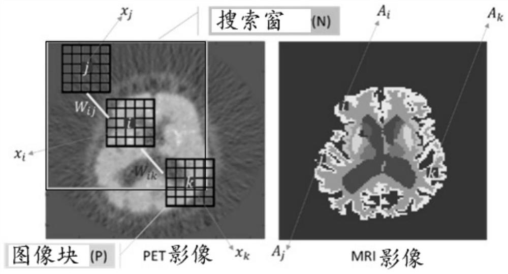
[0052] Step (4) introducing the non-local mean prior of MR image constraints into the PET partial volume correction model to construct a PET partial volume correction model based on NLMA;

[0053] In step (5), the objective function obtained in step (4) is iterativel...

Embodiment 2

[0081] A non-local mean-guided PET image partial volume correction method constrained by MR structure information, which is corrected according to the method described in Embodiment 1, the non-local mean-guided PET image partial volume correction method constrained by MR structure information.

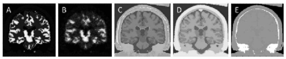
[0082] image 3 is the simulated image data used in the experiment in Example 2, which is obtained from real human body PET data, where image 3 (a) is the PET ideal image corresponding to the real object, the size is 128*128. image 3 (c) is the MR simulation image, which is divided into 6 different brain regions. image 3 (d) MR simulation image with segmentation error obtained with the SPM12 tool, also containing 6 different brain regions.

[0083] For the PET simulation image, the uncorrected PET image is obtained by MLEM iteration 240 times after attenuation correction, normalization correction, and photon count reduction in the projection domain, such as image 3 (b) shown. F...

Embodiment 3

[0089] A non-local mean-guided PET image partial volume correction method constrained by MR structure information, other features are the same as those in Embodiment 1 or 2, the difference is that the verification is performed according to Embodiment 2, and the method of the present invention is verified in order to verify the MR image segmentation deviation The influence of the two kinds of MR segmentation images is compared as the structure prior experiment, and the related results are as follows.

[0090] For PET simulation images, uncorrected PET images are obtained by attenuation correction, normalization correction, and photon count reduction in the projection domain, such as image 3 (b) shown. The RBV algorithm extends the GTM algorithm to pixels, is an algorithm based on MR segmentation, and is also a typical algorithm for partial volume correction using MR information. In this implementation 2, this method is selected for comparison with the method of the present inv...


PUM


Abstract

A non-local mean-guided PET image partial volume correction method constrained by MR structure information. The NLM search range is constrained by the MR structure information. While using the MR structure information, it can avoid the generated PET image from affecting the MR image. Over-reliance, improve PET image quality. The present invention uses MR structural information to correct the partial volume of PET images, improves the accuracy and efficiency of partial volume correction of PET images, uses the structural region of MR to constrain the search window of NLM, and reduces the excessive dependence of PET correction result images on MR images , to improve the accuracy of PET-corrected images.

Description

technical field [0001] The invention relates to the technical field of medical image processing, in particular to a PET image partial volume correction method guided by non-local mean value constrained by MR structure information. Background technique [0002] Positron emission tomography (PET) is an important imaging tool for clinical diagnosis and research at the molecular level. Due to the insufficient spatial resolution of the detector, the partial volume effect is more pronounced compared with MRI / CT imaging equipment. The partial volume effect will blur the image and distort the lesion, leading to the degradation of image quality and affecting clinical diagnosis and quantitative evaluation. PET partial volume correction methods can be divided into two categories: correction during reconstruction and correction during post-reconstruction. Each type of method can be further divided into voxel-level and region-of-interest-level correction methods. [0003] Partial volu...

Claims


Application Information

Patent Timeline
Patent Type & Authority Patents(China)
IPC IPC(8): G06T5/00G06T7/33
CPCG06T5/002G06T2207/10088G06T2207/10104G06T7/33
Inventor 高园园路利军阳维冯前进
Owner SOUTHERN MEDICAL UNIVERSITY
Why Eureka
Try Eureka