Supercharge Your Innovation With Domain-Expert AI Agents!

3D model construction and preparation method for mitral regurgitation and calcification stenosis

A mitral valve and model technology, applied in the field of 3D printing, can solve problems such as subvalvular structural limitations, and achieve the effect of reducing operation time and radiation intake

Active Publication Date: 2020-06-05
西安马克医疗科技有限公司
View PDF11 Cites 1 Cited by
  • Summary
  • Abstract
  • Description
  • Claims
  • Application Information

AI Technical Summary

Problems solved by technology

[0005] The purpose of the present invention is to provide a method for constructing and preparing a 3D model for mitral regurgitation and calcified stenosis, so as to solve the problems of traditional imaging assessment of mitral valve structure and subvalvular structure limitations in the prior art.

Method used


Image

  • 3D model construction and preparation method for mitral regurgitation and calcification stenosis
  • 3D model construction and preparation method for mitral regurgitation and calcification stenosis
  • 3D model construction and preparation method for mitral regurgitation and calcification stenosis

Examples


Embodiment 1

[0060] Disclosed in this embodiment is a method for constructing a 3D model for mitral valve disease, which is implemented in the Mimics software using the following steps:

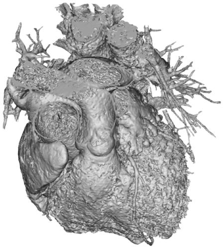
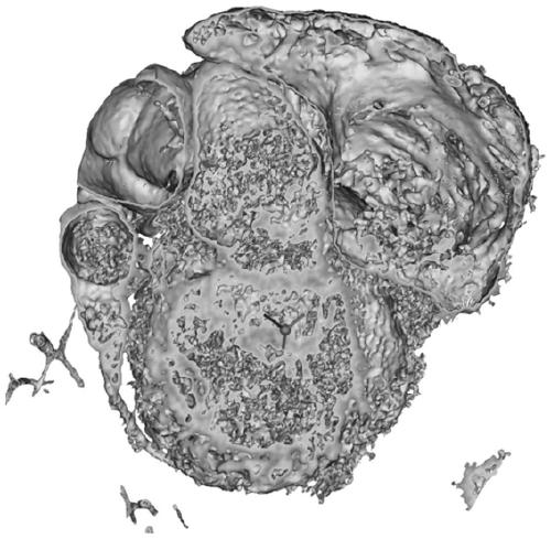
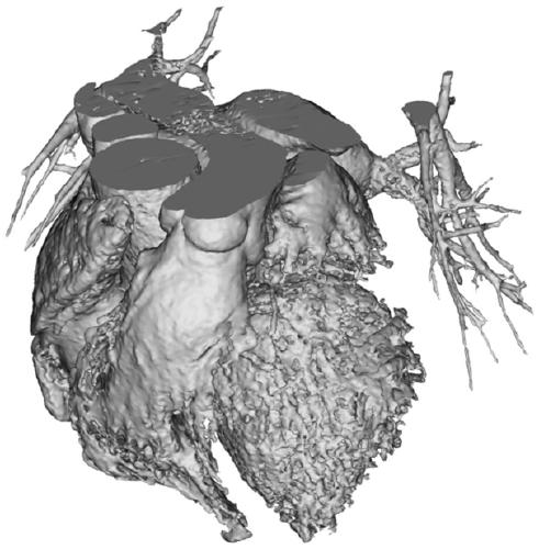
[0061] (1) Use medical imaging equipment to collect data on patients with mitral valve disease (calcified mitral valve stenosis, mitral regurgitation) left heart angiography, mainly collecting the patient's aortic root, part of the ascending aorta, coronary artery and left Atrium, left ventricle, and generate CT (DICOM) files including systole and diastole.

[0062] (2) Import the DICOM file into Mimics software, and generate a .mcs file to save

[0063] (3) Split data:

[0064] Step1, in the Pseudo colors command, according to the different chromatograms displayed by different tissues, observe the structure and boundary of different tissues, calcification, leaflet shape and degree of calcification;

[0065] Step2, CT image playback selects the phase data when the left heart is at its maximum or minimum...

Embodiment 2

[0074] The software selected for printing in this embodiment is OBJET slicing software, and slicing software such as FDM Cura, SLAMaterialise magics, and SLM QuantAM can also be selected.

[0075] This embodiment discloses a method for preparing a 3D model for mitral valve regurgitation and calcified stenosis. On the basis of Embodiment 1, it also includes the following steps:

[0076] Step 1: Use any of the above-mentioned 3D model construction methods for mitral regurgitation and calcification stenosis to obtain a 3D model for mitral regurgitation and calcification stenosis, and export the model as an STL format file;

[0077] Step 2: Import the 3D model into the OBJET slicing software for printing. During the slicing process, the height of the model should be as low as possible to reduce the printing time. After removing the large support on the surface of the model with tools, place the printed 3D model in alkaline Shake and clean in the solution. After cleaning, take the ...


PUM


Abstract

The invention belongs to the field of 3D printing, and discloses a 3D model construction and preparation method for mitral regurgitation and calcification stenosis. In the model construction method, firstly, left heart structure radiography data of a mitral regurgitation and calcification stenosis patient are acquired and preprocessed to obtain heart CT data; then, a calcification tissue model anda blood model are established according to gray values corresponding to different tissue in the heart CT data; valve leaflet chordae tendineae and papillary muscle are reconstructed, extra artifact blood structures in an inner cavity model are deleted to obtain the inner cavity model; and finally, the inner cavity model is cut, the cut inner cavity model and the calcification tissue model are subjected to Boolean operation to obtain a 3D model for mitral calcification stenosis and mitral regurgitation diseases. The 3D model construction and preparation method well solves the problems of limitation of traditional image evaluation mitral structures and structures under mitral valves in the prior art.

Description

technical field [0001] The invention belongs to the field of 3D printing, and in particular relates to a method for constructing and preparing a 3D model for mitral valve regurgitation and calcified stenosis. Background technique [0002] Mitral valve disease is the most common valvular disease that endangers the cardiovascular health of the population. The vast majority of mitral stenosis (Mitral stenosis, MS) is the sequelae of rheumatic fever, very few are congenital stenosis or senile mitral valve ring or subannular calcification. Mitral insufficiency (MI) is mainly caused by valvular degeneration, rheumatic fever, structural damage of the mitral valve subvalvular after coronary heart disease myocardial infarction, and often causes atrial fibrillation and cardiac insufficiency. According to the latest epidemiological data of western developed countries such as the United States, mitral valve regurgitation is the most common type of valvular disease among the elderly ove...

Claims


Application Information

Patent Timeline
IPC IPC(8): A61B34/10B33Y10/00
CPCA61B34/10B33Y10/00A61B2034/102
Inventor 李亚杰曾博文马克军
Owner 西安马克医疗科技有限公司
Features
  • R&D
  • Intellectual Property
  • Life Sciences
  • Materials
  • Tech Scout
  • Unparalleled Data Quality
  • Higher Quality Content
  • 60% Fewer Hallucinations
Learn More