3D model construction method and preparation method for aortic valve disease

An aortic valve and construction method technology, applied in the field of 3D printing, can solve the problems of misleading preoperative evaluation of the model, low accuracy of the aortic valve leaflet model, etc., to improve the surgical effect, high accuracy, and ensure production accuracy. Effect

Active Publication Date: 2020-06-05
西安马克医疗科技有限公司
View PDF12 Cites 4 Cited by
  • Summary
  • Abstract
  • Description
  • Claims
  • Application Information

AI Technical Summary

Problems solved by technology

[0005] The purpose of the present invention is to provide a 3D model construction method and preparation method for aortic valve disease, which is used to solve the problem that the reconstru

Method used


Image

  • 3D model construction method and preparation method for aortic valve disease
  • 3D model construction method and preparation method for aortic valve disease
  • 3D model construction method and preparation method for aortic valve disease

Examples


Embodiment 1

[0062] Disclosed in this embodiment is a method for building a 3D model for aortic valve disease, which is implemented in the Mimics software using the following steps:

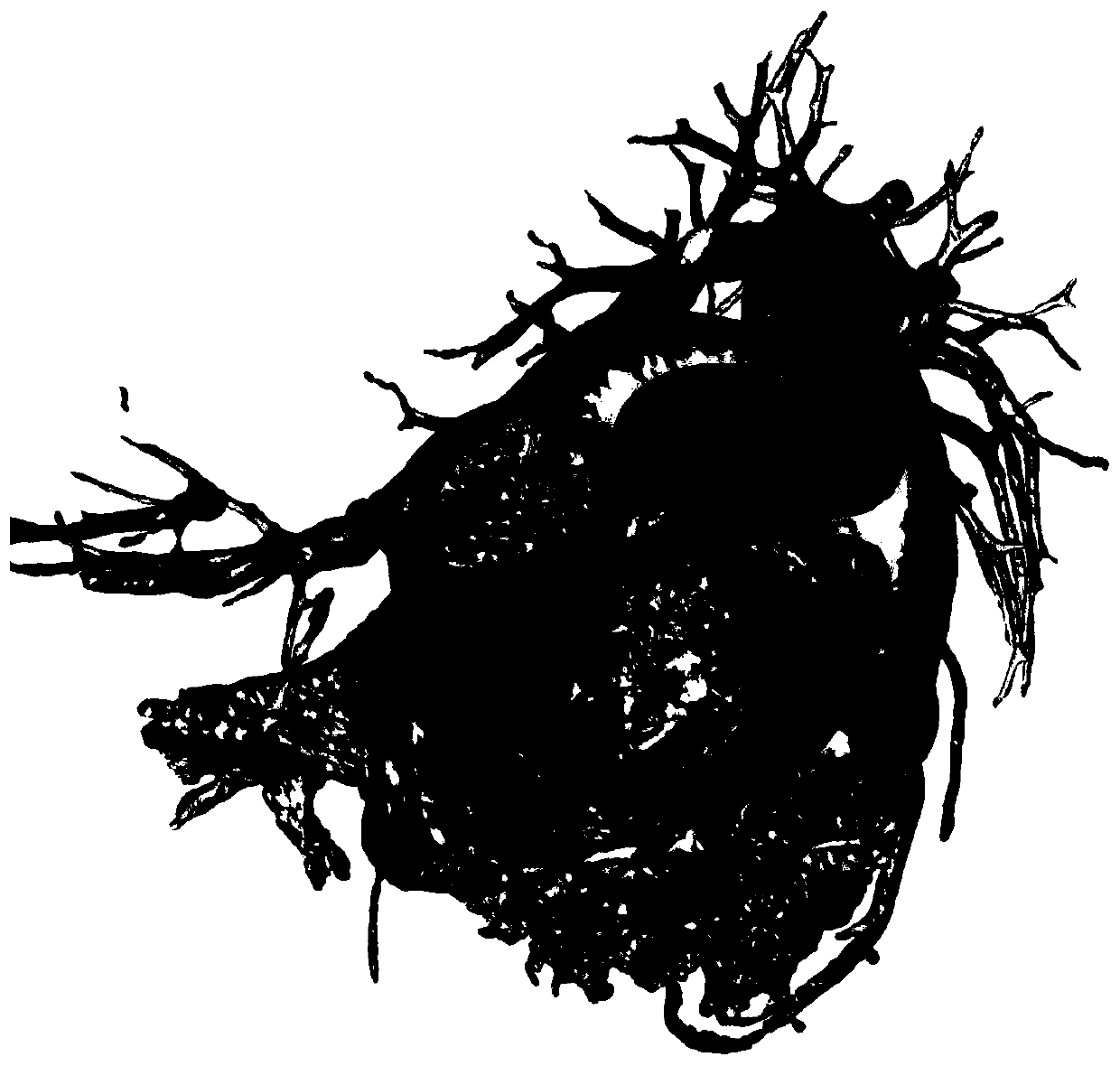
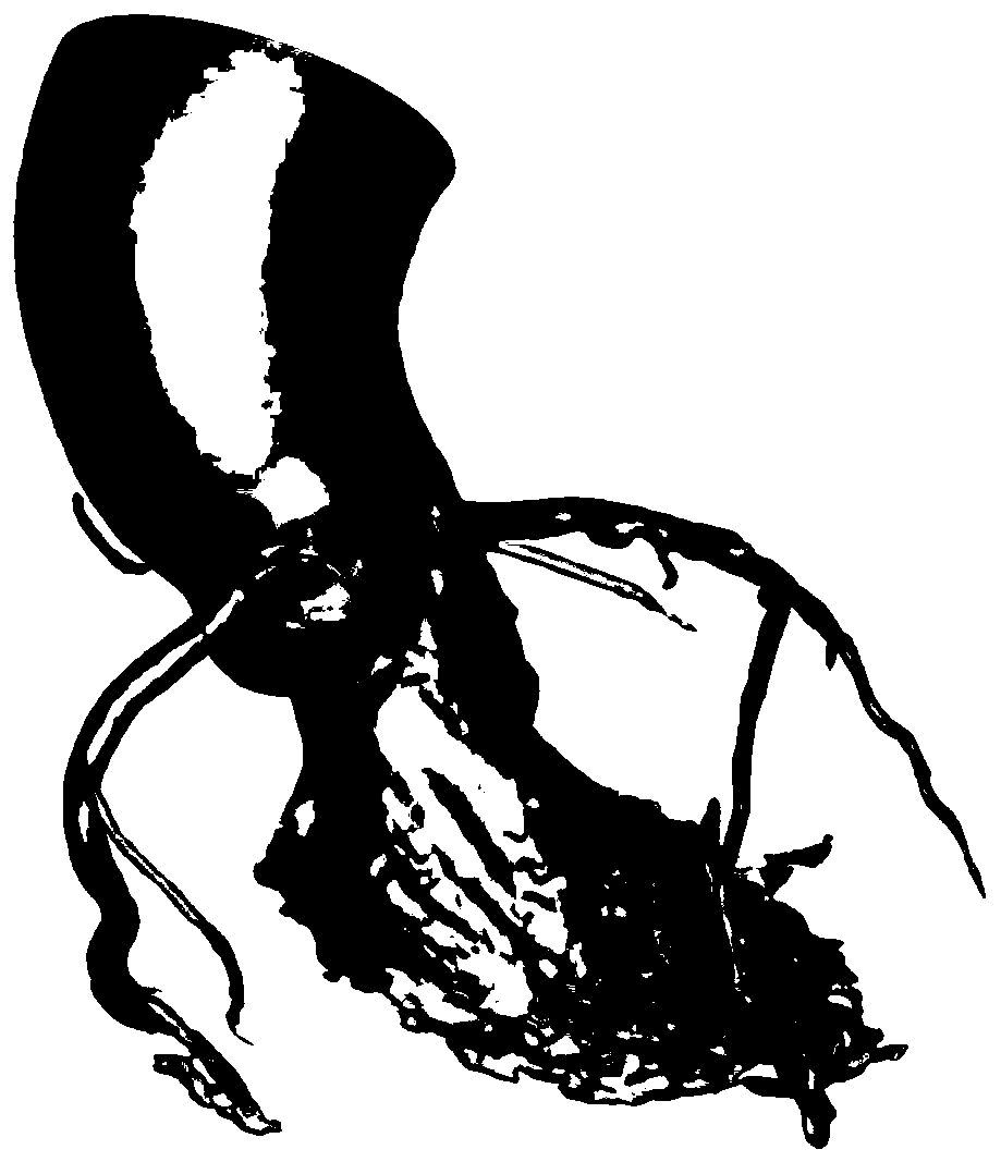
[0063] (1) Use medical imaging equipment to collect data on aortic angiography of patients with aortic valve disease (aortic valve stenosis, aortic valve insufficiency, aortic valve stenosis), mainly collecting the patient's aortic root and part of the ascending aorta , coronary artery and left atrium, and generate CT (DICOM) files including systole and diastole.

[0064] (2) Import the DICOM file into Mimics software, and generate a .mcs file to save

[0065] (3) Split data:

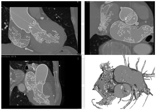
[0066] Step1, in the Pseudo colors command, according to the different chromatograms displayed by different tissues, observe the structure and boundary of different tissues, calcification, leaflet shape and degree of calcification;

[0067] Step2, CT image playback selects the phase data with the largest leaflet opening, and adjusts ...

Embodiment 2

[0076] This embodiment discloses a method for preparing a 3D model for aortic valve disease. On the basis of Embodiment 1, it also includes the following steps:

[0077] Step 1: Use any of the above-mentioned 3D model construction methods for aortic valve disease to obtain a 3D model for aortic valve disease, and export the model as an STL format file;

[0078] Step 2: Import the 3D model into the OBJET slicing software for printing. During the slicing process, the height of the model should be as low as possible to reduce the printing time. After removing the large support on the surface of the model with tools, place the printed 3D model in alkaline Shake and clean in the solution. After cleaning, take the model out of the solution and rinse the surface residue. Use a water gun tool to rinse the residual support material and alkaline solution on the surface;

[0079] Step 3: Put the 3D model obtained in step 2 into the drying box and keep the blast mode for drying, take out ...


PUM


Abstract

The invention belongs to the field of 3D printing and discloses a 3D model construction method for an aortic valve disease. The 3D model construction method comprises the following steps: step 1: acquiring and preprocessing aorta radiography data of an aortic valve disease patient to obtain heart CT data; step 2: establishing a calcified tissue model and a blood model according to gray values corresponding to different tissue in the heart CT data; step 3: comparing a valve leaflet border with an actual valve leaflet border and correction border in the heart CT data to obtain an inner cavity model; and step 4: deleting blood and reserving an aortic root, a partial coronary artery and a left ventricle outflow tract to obtain a 3D model for the aortic valve disease. The invention also discloses a 3D model preparation method for the aortic valve disease. The model construction efficiency is high, all that is required is to reconstruct the blood form in a blood vessel cavity and then rapidly segmented required tissue, whole reconstruction of blood vessel walls and other hearts and then segmentation are not needed, and a lot of time is saved for model repair.

Description

technical field [0001] The invention belongs to the field of 3D printing, and in particular relates to a 3D model construction method and preparation method for aortic valve disease. Background technique [0002] Aortic valve disease is the functional or structural abnormality of single or multiple valve structures caused by inflammation, myxoid degeneration, degenerative changes, etc., resulting in valve stenosis and (or) insufficiency. Aortic valve calcification is a kind of congenital aortic valve calcification, which is mainly caused by the reduction of blood vessel elasticity or high blood pressure with age; in recent years, with the improvement of economic conditions and medical level, human The life expectancy is prolonged, and the incidence rate is significantly increased. Aortic valve calcification has become a prominent problem in geriatric clinical cardiology. [0003] Patients with mild aortic valve calcification can be controlled by drugs, but more severe patie...

Claims


Application Information

Patent Timeline
IPC IPC(8): A61B34/10B33Y10/00
CPCA61B34/10B33Y10/00A61B2034/102
Inventor 李亚杰曾博文马克军
Owner 西安马克医疗科技有限公司
Why Eureka
Try Eureka