Improved DME edema area neural network segmentation model construction method

A neural network and segmentation model technology, which is applied in the field of neural network segmentation model construction in DME edema area, can solve problems affecting the calculation accuracy of the segmentation model, focusing on pathological classification, increasing neural network training time, etc.

Active Publication Date: 2020-06-26
SHANGHAI OCEAN UNIV +1
View PDF4 Cites 0 Cited by
  • Summary
  • Abstract
  • Description
  • Claims
  • Application Information

AI Technical Summary

Problems solved by technology

Haeker et al. regarded the OCT image as a graph model, and the pixel value as a node, and segmented the edema part based on graph theory, but using pixels as nodes increased the training time of the neural network, and the sensitivity of the segmentation results was low; Roy et al. A retinal segmentation algorithm based on the U-net neural network is proposed, but the shallow network cannot extract high-dimensional abstract features, and the segmentation accuracy is low on complex images with partial pathological coexistence; Kermany et al. implemented a multi- Eye disease detection method, but this model focuses on pathological classification and cannot accurately segment the lesion
[0005] To sum up, there are several problems in the existing technology of using OCT images to quickly and accurately segment DME edema areas: (1) Multi-source OCT The heterogeneity of the image affects the calculation accuracy of the segmentation model: it is affected by factors such as the equipment that produces the OCT image, the level of the operator, the external environment, and the pathogen itself; there is a large heterogeneity in the OCT image, and the quality of some OCT images is low. Phenomena such as speckle noise and mechanical noise exist (such as Figure 9shown)
At the same time, the visual interpretation of the DME edema area has problems such as time-consuming and unstable accuracy.
[0007](3) The ambiguity of the boundary of the DME area poses a challenge to the practicability of the segmentation model: the coexistence of pathology will make the overall appearance of the lesion complex, how to obtain High-precision lesion region boundary is a problem to be solved by the segmentation model
[0008](4) In the existing automatic segmentation method of DME region based on machine learning and deep learning, the edema part is segmented based on graph theory, but the pixel The training time of the neural network is increased for the nodes, and the sensitivity of the segmentation results is low; the shallow network cannot extract high-dimensional abstract features, and the segmentation accuracy is low on complex images with some pathological coexistence; the existing model focuses on pathological classification and cannot Accurate segmentation of lesion
[0009]Difficulty in solving the above technical problems: the influence of multiple sources of OCT images, the diversity of DME region features, and the blurring of DME region boundaries, the visual inspection of DME edema region Interpretation has problems such as time-consuming and unstable accuracy

Method used


Image

  • Improved DME edema area neural network segmentation model construction method
  • Improved DME edema area neural network segmentation model construction method
  • Improved DME edema area neural network segmentation model construction method

Examples


Embodiment 1

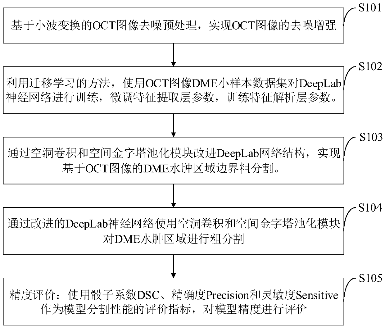
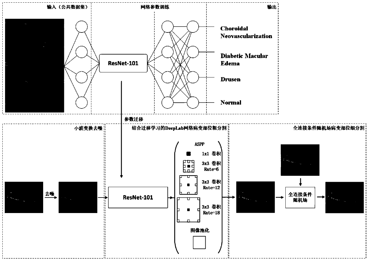
[0095] Such as figure 2 As shown, the improved DME edema area neural network segmentation model construction method provided by the embodiment of the present invention specifically includes:

[0096] 1.1 OCT image enhancement based on wavelet transform

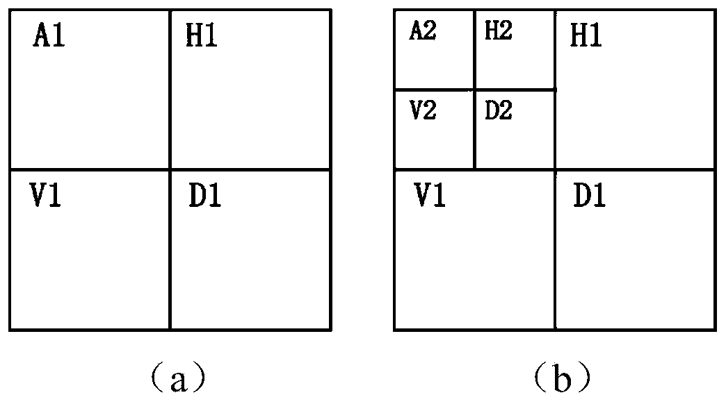
[0097] OCT images have the characteristics of image noise and unclear lesion area, which affect the segmentation accuracy of DME edema area. Wavelet transform can denoise OCT images. Use wavelet transform to decompose the OCT image, and decompose the OCT image into a low-frequency sub-band and three high-frequency sub-bands, in which noise is mostly distributed in the low-frequency sub-band; edge and texture information are mostly distributed in the three high-frequency sub-bands; Through decomposition, the low-frequency sub-bands can be processed separately, and the noise in the low-frequency sub-bands can be removed without affecting the edge and texture information of the high-frequency sub-bands. If one-time decomposit...

Embodiment 2

[0123] 2.1 Accuracy evaluation

[0124] Using the dice coefficient DSC, precision Precision, and sensitivity Sensitive as the evaluation index of model segmentation performance, the calculation formula of each system is as follows:

[0125]

[0126]

[0127]

[0128] Among them, Vs and Vg represent the lesion area obtained by model segmentation and the lesion area obtained by visual interpretation, respectively.


PUM


Abstract

The invention belongs to the technical field of network data processing, and discloses an improved DME edema area neural network segmentation model construction method, which comprises the steps of performing OCT image denoising preprocessing; based on an improved DeepLab neural network, achieving coarse segmentation of a DME edema area, and designing a DeepLab neural network structure by using hole convolution and a spatial pyramid pooling module; introducing a full-connection conditional random field to optimize a DME edema region boundary; and evaluating the segmentation model precision byutilizing the evaluation indexes of the model segmentation performance. According to the method, the contrast of the image can be improved, the edge texture information of the lesion part is reservedwhile the noise is removed, and a good image data foundation is laid for accurate identification and segmentation of the edema area; good lesion part segmentation performance can be obtained, the feeling visual field is enlarged, the segmentation performance is enhanced, and the segmentation speed of the OCT image is improved.

Description

technical field [0001] The invention belongs to the technical field of neural network segmentation model construction in DME edema region, relates to an improved method for constructing a neural network segmentation model in DME edema region, in particular to an improved DME edema region neural network combined with wavelet transform and fully connected conditional random field Split the model. Background technique [0002] At present, diabetic retinopathy is the main complication of diabetes in the eyes, and it is one of the four major blindness diseases in my country. With the improvement of people's living standards in our country, the incidence and blindness rate of diabetic retinopathy have increased significantly in recent years, which seriously affects the visual function and quality of life of patients. Diabetic macular edema (DME) is a common cause of visual impairment in diabetic patients. DME mainly refers to retinal thickening or hard exudative deposits caused b...

Claims


Application Information

Patent Timeline
IPC IPC(8): G06T7/12G06T5/10G06T5/00
CPCG06T7/12G06T5/10G06T2207/10101G06T2207/20016G06T2207/20081G06T2207/20084G06T2207/20064G06T2207/30041G06T2207/30096G06T5/70
Inventor 王振华钟元芾蒋沁李超鹏颜标李秀苗姚牧笛
Owner SHANGHAI OCEAN UNIV
Why Eureka
Try Eureka